File:PMC3679835 1869-5760-3-49-3.png
Jump to navigation
Jump to search
No higher resolution available.
PMC3679835_1869-5760-3-49-3.png (512 × 187 pixels, file size: 164 KB, MIME type: image/png)
File history
Click on a date/time to view the file as it appeared at that time.
| Date/Time | Thumbnail | Dimensions | User | Comment | |
|---|---|---|---|---|---|
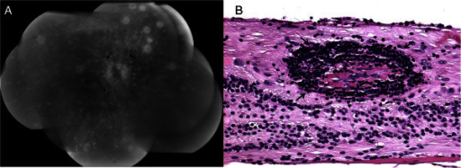
| current | 22:37, 11 January 2022 | 512 × 187 (164 KB) | Ozzie10aaaa | Author:Chu XK, Chan CC, Immunopathology Section, Laboratory of Immunology, National Eye Institute, National Institutes of Health(Openi?national Library of Medicine) Source:https://openi.nlm.nih.gov/detailedresult?img=PMC3679835_1869-5760-3-49-3&query=Sympathetic%20ophthalmia&it=xg&req=4&npos=1 Description:F3: Visualization of mild retinal vasculitis, an uncommon finding in sympathetic ophthalmia. (A) Fluorescein angiogram of a sympathetic ophthalmia retina shows mild vascular leakage revealin... |
File usage
There are no pages that use this file.