File:PMC3493364 1471-2474-13-132-2.png
Jump to navigation
Jump to search
No higher resolution available.
PMC3493364_1471-2474-13-132-2.png (512 × 384 pixels, file size: 381 KB, MIME type: image/png)
File history
Click on a date/time to view the file as it appeared at that time.
| Date/Time | Thumbnail | Dimensions | User | Comment | |
|---|---|---|---|---|---|
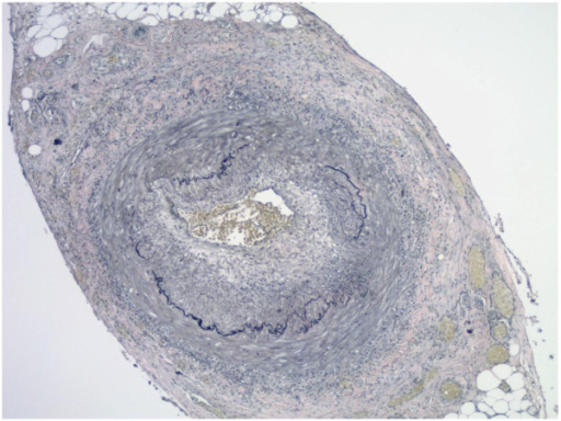
| current | 21:54, 27 March 2022 | | 512 × 384 (381 KB) | Ozzie10aaaa | Author:Mohammadi A, Pfeifer JD, Lewis JS , Department of Pathology and Laboratory Medicine, University of Florida, College of Medicine(Openi/National Library of Medicine)Source:https://openi.nlm.nih.gov/detailedresult?img=PMC3493364_1471-2474-13-132-2&query=temporal%20arteritis&it=xg&req=4&npos=2 Description:F2: Elastic stains on the same case of histologically positive temporal artery biopsy as shown in Figure1, showing the fragmentation, distortion and lack of continuity of the internal ela... |
File usage
The following page uses this file: