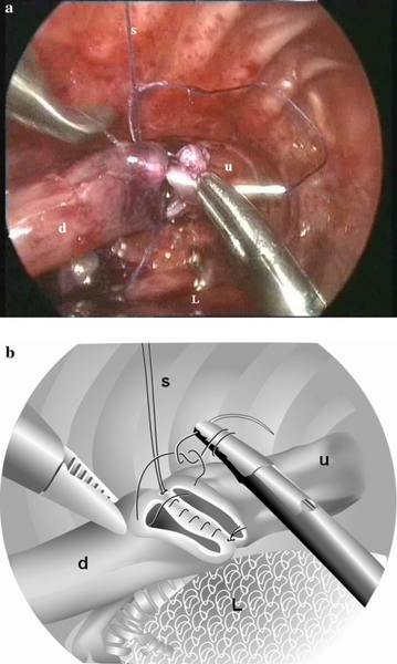
File:PMC3414695 268 2012 1651 Fig2 HTML.png
Jump to navigation
Jump to search

Size of this preview: 359 × 600 pixels. Other resolutions: 144 × 240 pixels | 512 × 855 pixels.
Original file (512 × 855 pixels, file size: 632 KB, MIME type: image/png)
File history
Click on a date/time to view the file as it appeared at that time.
| Date/Time | Thumbnail | Dimensions | User | Comment | |
|---|---|---|---|---|---|
| current | 20:38, 2 January 2023 |  | 512 × 855 (632 KB) | Ozzie10aaaa | Uploaded a work by van der Zee DC, Tytgat SH, Zwaveling S, van Herwaarden MY, Vieira-Travassos D from https://openi.nlm.nih.gov/detailedresult?img=PMC3414695_268_2012_1651_Fig2_HTML&query=Esophageal%20atresia&it=xg&req=4&npos=9 with UploadWizard |
File usage
There are no pages that use this file.