File:PMC3362223 cop-0003-0142-g01 (1).png
Jump to navigation
Jump to search
No higher resolution available.
PMC3362223_cop-0003-0142-g01_(1).png (512 × 246 pixels, file size: 300 KB, MIME type: image/png)
.png)
File history
Click on a date/time to view the file as it appeared at that time.
| Date/Time | Thumbnail | Dimensions | User | Comment | |
|---|---|---|---|---|---|
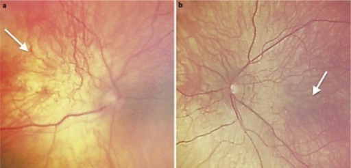
| current | 00:11, 30 January 2022 | | 512 × 246 (300 KB) | Ozzie10aaaa | Author:Wang BZ, Siriwardana P, Taranath D , Department of Ophthalmology, Flinders Medical Centre and Flinders University(Openi/National Library of Medicine) Source:https://openi.nlm.nih.gov/detailedresult?img=PMC3362223_cop-0003-0142-g01&query=Macular%20hypoplasia&it=xg&req=4&npos=1 Description:F1: Fundus photographs of the right eye (a) and left eye (b) showing macular hypoplasia (white arrows) with blond retinal background and underlying choroidal vessels visible. |
File usage
There are no pages that use this file.
.png)