File:PMC3309207 arm-35-436-g003.png
Jump to navigation
Jump to search
No higher resolution available.
PMC3309207_arm-35-436-g003.png (471 × 196 pixels, file size: 178 KB, MIME type: image/png)
File history
Click on a date/time to view the file as it appeared at that time.
| Date/Time | Thumbnail | Dimensions | User | Comment | |
|---|---|---|---|---|---|
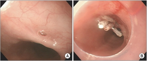
| current | 23:03, 7 January 2022 | | 471 × 196 (178 KB) | Ozzie10aaaa | Author:Jung JH, Kim JS, Kim YK,Department of Physical Medicine and Rehabilitation, Kwandong University College of Medicine(Openi/National Library of Medicine) Source:https://openi.nlm.nih.gov/detailedresult?img=PMC3309207_arm-35-436-g003&query=Tracheoesophageal%20fistula&it=xg&req=4&npos=5 Description:F3: Esophagoduodenoscopic images: About the 3 mm-sized tracheoesophageal fistula was seen. And also the bubble from the trachea was observed. The fistula was located in the esophagus about 33 cm... |
File usage
There are no pages that use this file.