File:PMC3271596 JNRP-3-100b-g001.png
Jump to navigation
Jump to search
No higher resolution available.
PMC3271596_JNRP-3-100b-g001.png (512 × 168 pixels, file size: 127 KB, MIME type: image/png)
File history
Click on a date/time to view the file as it appeared at that time.
| Date/Time | Thumbnail | Dimensions | User | Comment | |
|---|---|---|---|---|---|
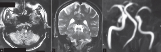
| current | 23:45, 1 February 2022 | 512 × 168 (127 KB) | Ozzie10aaaa | Author:Shetty SR, Anusha R, Thomas PS, Babu SG, Department of Oral Medicine and Radiology, AB Shetty Memorial Institute of Dental Sciences, Nitte University(Openi/National Library of Medicine)Source:https://openi.nlm.nih.gov/detailedresult?img=PMC3271596_JNRP-3-100b-g001&query=Wallenberg%20syndrome&it=xg&req=4&npos=3 Description:F1: (a) A FLAIR image showing a high signal in the left postero lateral medulla suggestive of an infarct (circle in image) and there is a white round lesion immediate... |
File usage
There are no pages that use this file.