File:PMC3078321 431 2011 1452 Fig1 HTML.png
Jump to navigation
Jump to search
No higher resolution available.
PMC3078321_431_2011_1452_Fig1_HTML.png (496 × 372 pixels, file size: 256 KB, MIME type: image/png)
File history
Click on a date/time to view the file as it appeared at that time.
| Date/Time | Thumbnail | Dimensions | User | Comment | |
|---|---|---|---|---|---|
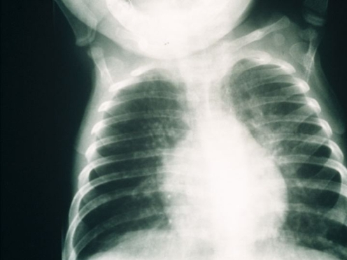
| current | 23:16, 6 February 2022 | | 496 × 372 (256 KB) | Ozzie10aaaa | Author:van der Burg M, Gennery AR, Department of Immunology, Erasmus MC, University Medical Center Rotterdam(Openi/National Library of Medicine) Source:https://openi.nlm.nih.gov/detailedresult?img=PMC3078321_431_2011_1452_Fig1_HTML&query=Severe%20combined%20immunodeficiency&it=xg&req=4&npos=1 Description:Fig1: Chest radiograph from a 5-month-old infant with severe combined immunodeficiency showing bilateral patchy shadowing secondary to interstitial pnuemonitis due to infection with respirato... |
File usage
The following page uses this file: