No higher resolution available.
This file is from a shared repository and may be used by other projects.
The description on its file description page there is shown below.
License
Attribution 3.0 Unported (CC BY 3.0)
Summary
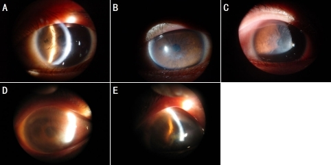
Author:Zhang C, Zeng G, Lin H, Li D, Zhao L, Zhou N, Qi Y, Department of Ophthalmology, Harbin Medical University the 2nd Affiliated Hospital (Openi/National Library of Medicine) Source:https://openi.nlm.nih.gov/detailedresult?img=PMC2786890_mv-v15-2498-f2&query=Secondary%20systemic%20amyloidosis&it=xg&req=4&npos=2 Description:f2: Clinical photographs of affected family members with LCD I. A and B: Slit lamp appearance of a cornea of a proband at 16 years of age shows distinct refractile lattice lines and diffuse opacification in the subepithelial and anterior stromal layer in the left eye. C: The photograph demonstrates central anterior stromal clouding in the proband’s right eye. D: The photograph shows lattice opacities with the appearance of new vessels in the subepithelial and anterior stromal cornea of the proband’s mother, with recurrent disease in the right eye. E: The image of the proband’s mother reveals thick linear opacities and diffuse grayish-white clouding, which partly covers the original lattice lines within the central area of the cornea in the left eye.
File history
Click on a date/time to view the file as it appeared at that time.
| Date/Time | Thumbnail | Dimensions | User | Comment |
|---|
| current | 20:42, 30 October 2021 |  | 482 × 241 (138 KB) | Ozzie10aaaa | Author:Zhang C, Zeng G, Lin H, Li D, Zhao L, Zhou N, Qi Y, Department of Ophthalmology, Harbin Medical University the 2nd Affiliated Hospital (Openi/National Library of Medicine) Source:https://openi.nlm.nih.gov/detailedresult?img=PMC2786890_mv-v15-2498-f2&query=Secondary%20systemic%20amyloidosis&it=xg&req=4&npos=2 Description:f2: Clinical photographs of affected family members with LCD I. A and B: Slit lamp appearance of a cornea of a proband at 16 years of age shows distinct refractile latt... |
File usage
The following page uses this file: