No higher resolution available.
This file is from a shared repository and may be used by other projects.
The description on its file description page there is shown below.
License
Attribution-NonCommercial 3.0 Unported (CC BY-NC 3.0)
Summary
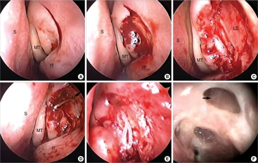
Author:Choi JC, Jin HR, Moon YE, Kim MS, Oh JK, Kim HA, Choi MY, Shim WS,Department of Otorhinolaryngology-Head and Neck surgery, Chungbuk National University(Openi/National Library of Medicine) Source:https://openi.nlm.nih.gov/detailedresult?img=PMC2751879_ceo-2-141-g002&query=Dacryocystorhinostomy&it=xg&req=4&npos=15 Description:F2: Operative technique of endoscopic dacryocystorhinostomy. (A) A vertical mucosal incision is made at the lateral nasal wall. (B) Mucosal flap is elevated and resected. (C) The maxillary bone covering the lacrimal sac is removed. (D) The anteriorly based lacrimal sac flap is everted and adjusted to accurately appose the nasal mucosa. Note that the common canalicular opening (indicated with arrow) is visible. (E) The silicone bicanalicular tube is positioned. (F) Nasal endoscopic finding six months after surgery. The rhinostomy opening (arrow) is wide and patent. S: septum; MT: middle turbinate; IT: inferior turbinate; LS: lacrimal sac.
File history
Click on a date/time to view the file as it appeared at that time.
| Date/Time | Thumbnail | Dimensions | User | Comment |
|---|
| current | 21:04, 3 February 2022 |  | 512 × 324 (423 KB) | Ozzie10aaaa | Author:Choi JC, Jin HR, Moon YE, Kim MS, Oh JK, Kim HA, Choi MY, Shim WS,Department of Otorhinolaryngology-Head and Neck surgery, Chungbuk National University(Openi/National Library of Medicine) Source:https://openi.nlm.nih.gov/detailedresult?img=PMC2751879_ceo-2-141-g002&query=Dacryocystorhinostomy&it=xg&req=4&npos=15 Description:F2: Operative technique of endoscopic dacryocystorhinostomy. (A) A vertical mucosal incision is made at the lateral nasal wall. (B) Mucosal flap is elevated and res... |
File usage
The following page uses this file: