File:PMC2621132 1757-1626-1-397-1.png
Jump to navigation
Jump to search
No higher resolution available.
PMC2621132_1757-1626-1-397-1.png (512 × 308 pixels, file size: 119 KB, MIME type: image/png)
File history
Click on a date/time to view the file as it appeared at that time.
| Date/Time | Thumbnail | Dimensions | User | Comment | |
|---|---|---|---|---|---|
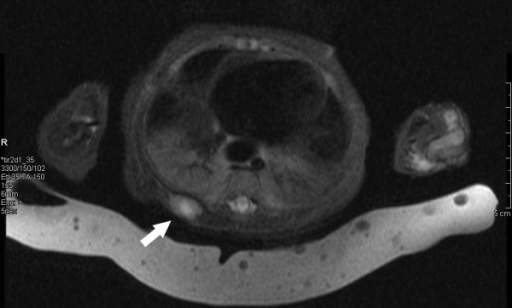
| current | 23:30, 22 February 2022 | | 512 × 308 (119 KB) | Ozzie10aaaa | Author:Dhall D, Frykman PK, Wang HL , Department of Pathology and Laboratory Medicine, Cedars-Sinai Medical Center(Openi/National Library of Medicine) Source:https://openi.nlm.nih.gov/detailedresult?img=PMC2621132_1757-1626-1-397-1&query=&req=4 Description:F1: Magnetic resonance imaging of the right posterior hemithorax showing a soft tissue lesion. The nodule was inferior to the level of the scapula and was within the paraspinal muscles measuring 1.5 × 0.9 × 1.8 cm. |
File usage
The following page uses this file: