File:Human Cortical Development.png
Jump to navigation
Jump to search
Size of this preview: 520 × 600 pixels. Other resolutions: 208 × 240 pixels | 416 × 480 pixels | 666 × 768 pixels | 888 × 1,024 pixels | 2,303 × 2,656 pixels.
Original file (2,303 × 2,656 pixels, file size: 1,022 KB, MIME type: image/png)
File history
Click on a date/time to view the file as it appeared at that time.
| Date/Time | Thumbnail | Dimensions | User | Comment | |
|---|---|---|---|---|---|
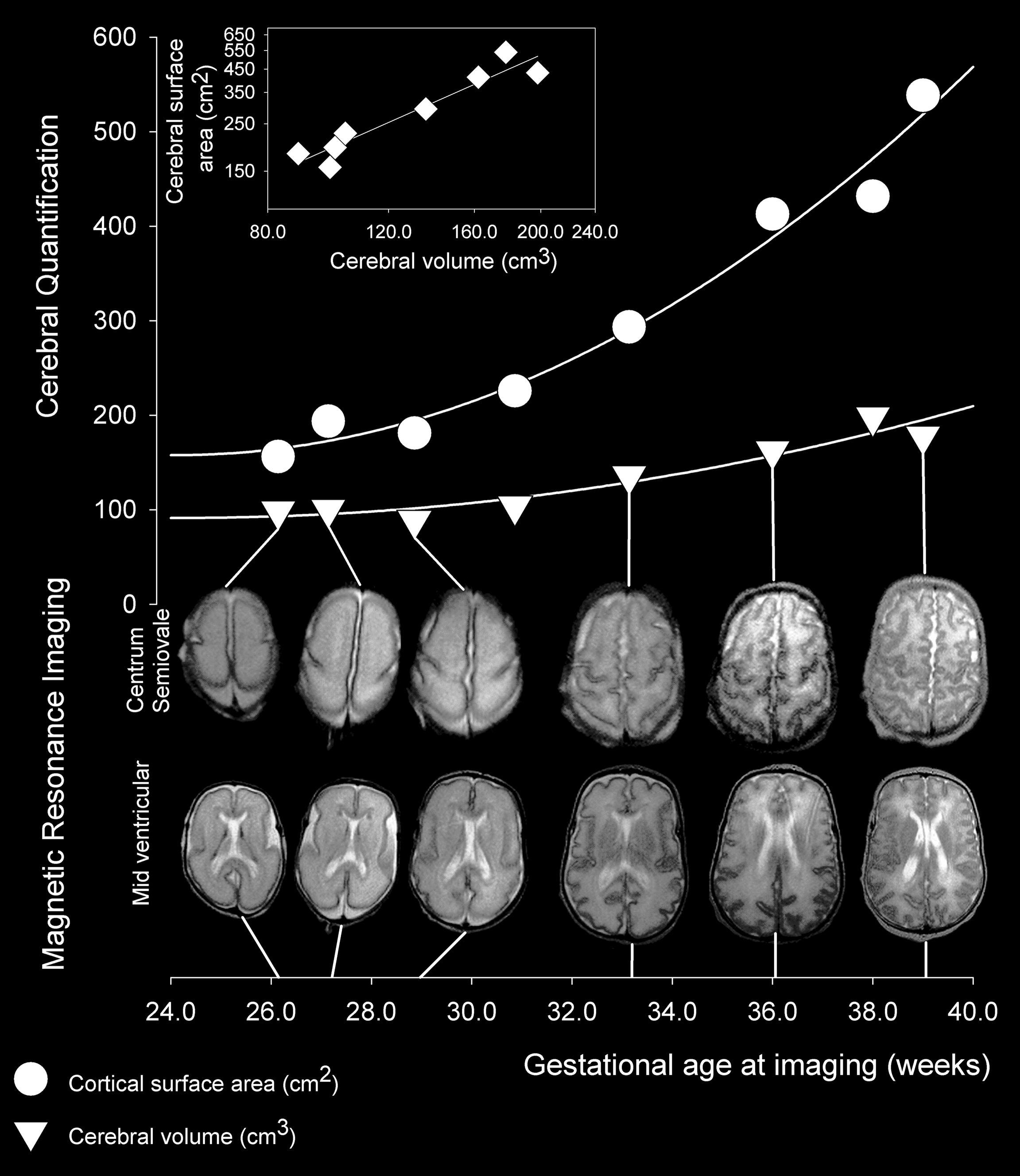
| current | 15:13, 5 January 2010 | | 2,303 × 2,656 (1,022 KB) | commons>Was a bee | {{Information |Description=The images show slices through the brain at the mid-ventricular level and at the level of the centrum semiovale from six of the eight MR images obtained between 26 and 39 week gestational age; images obtained at 30 and 38 weeks |
File usage
There are no pages that use this file.