File:Hepatocellular carcinoma (gross pathology) (Radiopaedia 8480).jpg
Jump to navigation
Jump to search
No higher resolution available.
Hepatocellular_carcinoma_(gross_pathology)_(Radiopaedia_8480).jpg (550 × 550 pixels, file size: 69 KB, MIME type: image/jpeg)
_(Radiopaedia_8480).jpg)
Summary:
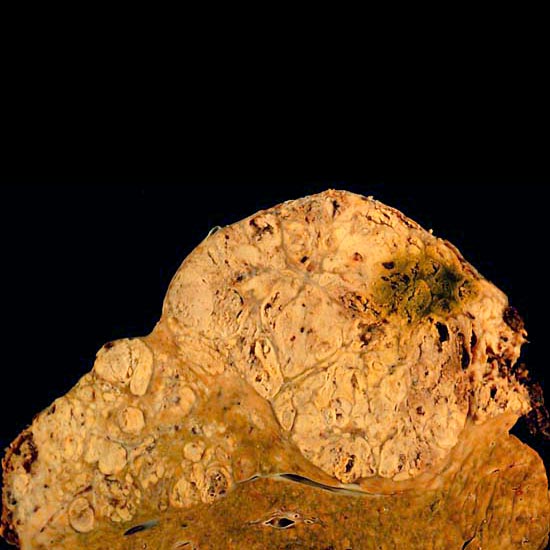
- Radiopaedia case ID: 8480
- Image ID: 260445
- Modality: Pathology
- System: Hepatobiliary
- Findings: The autopsy showed this hepatocellular carcinoma occupying much of the volume of a cirrhotic liver. Furthermore, the tumor had invaded the diaphragm and ruptured into the peritoneal cavity, causing the bloody ascites. The photo above shows a view of a longitudinal slice taken through the full length of the liver. The closer view, below, shows tumor at the top, cirrhotic liver at the bottom, and a fibrous reaction in between. Hepatocellular carcinomas can have a variety of gross patterns, including multinodular/multifocal, such as this one.
- Published: 4th Feb 2010
- Source: https://radiopaedia.org/cases/hepatocellular-carcinoma-gross-pathology-1
- Author: Ed Uthman
- Permission: http://creativecommons.org/licenses/by-nc-sa/3.0/
Licensing:
Attribution-NonCommercial-ShareAlike 3.0 Unported (CC BY-NC-SA 3.0)
| This file is ineligible for copyright and therefore in the public domain, because it is a technical image created as part of a standard medical diagnostic procedure. No creative element rising above the threshold of originality was involved in its production.
|
 |
File history
Click on a date/time to view the file as it appeared at that time.
| Date/Time | Thumbnail | Dimensions | User | Comment | |
|---|---|---|---|---|---|
| current | 17:00, 22 March 2021 | | 550 × 550 (69 KB) | Fæ | Radiopaedia project rID:8480 (batch #16294) |
File usage
The following page uses this file:
_(Radiopaedia_8480).jpg)