File:FSHDBodyDiagram.jpg
Jump to navigation
Jump to search
Size of this preview: 409 × 599 pixels. Other resolutions: 164 × 240 pixels | 328 × 480 pixels | 524 × 768 pixels | 1,265 × 1,852 pixels.
Original file (1,265 × 1,852 pixels, file size: 383 KB, MIME type: image/jpeg)
Summary
| Description |
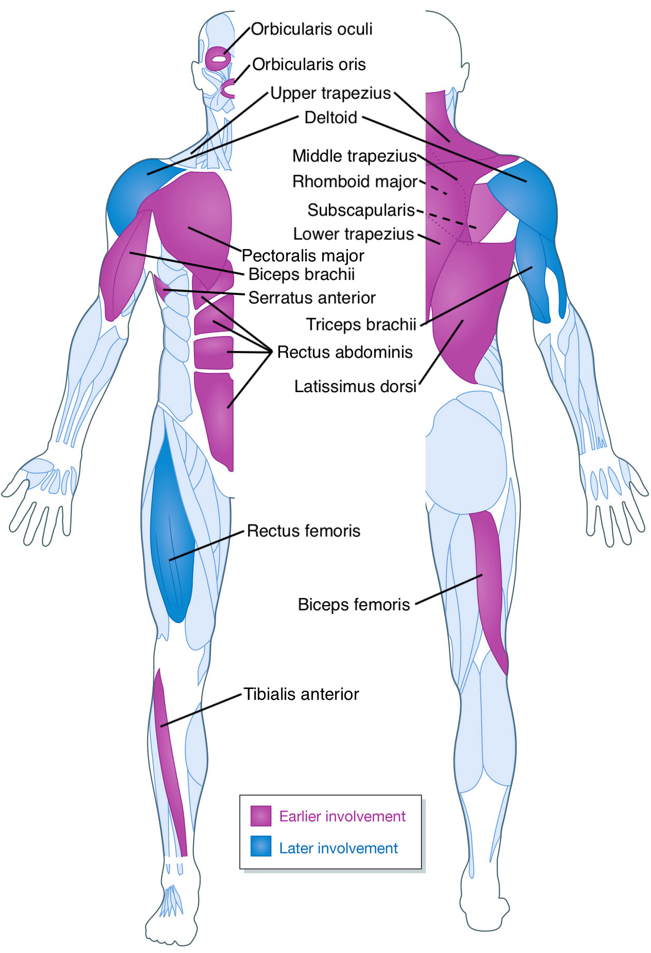
English: A diagram showing the muscles commonly involved in FSHD. Muscles most frequently involved are shown in pale purple, and those with later involvement shown in blue. |
| Date | |
| Source | Pathomechanisms and biomarkers in facioscapulohumeral muscular dystrophy: roles of DUX4 and PAX7. EMBO Mol Med (2021)e13695, https://pubmed.ncbi.nlm.nih.gov/34151531/ |
| Author |
Original author Daniel C. L. Zammit, used in publication by Christopher R S Banerji and Peter S Zammit edited by Lukelahood |
Licensing
This file is licensed under the Creative Commons Attribution 4.0 International license.
- You are free:
- to share – to copy, distribute and transmit the work
- to remix – to adapt the work
- Under the following conditions:
- attribution – You must give appropriate credit, provide a link to the license, and indicate if changes were made. You may do so in any reasonable manner, but not in any way that suggests the licensor endorses you or your use.
File history
Click on a date/time to view the file as it appeared at that time.
| Date/Time | Thumbnail | Dimensions | User | Comment | |
|---|---|---|---|---|---|
| current | 00:19, 7 July 2021 | | 1,265 × 1,852 (383 KB) | commons>Lukelahood | error corrected |
File usage
There are no pages that use this file.