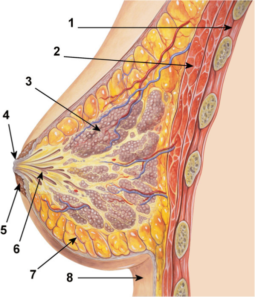
File:Diagram of the ductal anatomy of the breast.png
Jump to navigation
Jump to search
Diagram_of_the_ductal_anatomy_of_the_breast.png (512 × 596 pixels, file size: 518 KB, MIME type: image/png)
File history
Click on a date/time to view the file as it appeared at that time.
| Date/Time | Thumbnail | Dimensions | User | Comment | |
|---|---|---|---|---|---|
| current | 06:28, 26 January 2024 |  | 512 × 596 (518 KB) | Whispyhistory | Uploaded a work by Jütte, J., Hohoff, A., Sauerland, C. et al. from [https://bmcpregnancychildbirth.biomedcentral.com/articles/10.1186/1471-2393-14-124 In vivo assessment of number of milk duct orifices in lactating women and association with parameters in the mother and the infant]. BMC Pregnancy Childbirth 14, 124 (2014). https://doi.org/10.1186/1471-2393-14-124. [https://openi.nlm.nih.gov/detailedresult?img=PMC3992155_1471-2393-14-124-2&query=&req=4 Via Openi] with UploadWizard |
File usage
There are no pages that use this file.
