File:Breast cancer spheroids with aptamers.png
Jump to navigation
Jump to search
Size of this preview: 601 × 599 pixels. Other resolutions: 241 × 240 pixels | 481 × 480 pixels | 728 × 726 pixels.
Original file (728 × 726 pixels, file size: 613 KB, MIME type: image/png)
Summary
| Description |
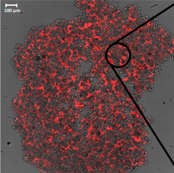
English: Breast cancer spheroids incubated with fluoresceinated SKBR3-R1 and MCF10A aptamers. Aptamers give off red light. Images show 2 levels of magnifigation (10x on the left, 100x in the center and right). The rightmost panel shows Hoescht-stained nuclei (cyan) as well as the red aptamer signal. |
| Date | |
| Source | Nelissen, F. H., Peeters, W. J., Roelofs, T. P., Nagelkerke, A., Span, P. N., & Heus, H. A. (2021). Improving breast cancer treatment specificity using aptamers obtained by 3D cell-SELEX. Pharmaceuticals, 14(4), 349. Publisher: MDPI. |
| Author | Frank H. T. Nelissen, Wenny J. M. Peeters, Timo P. Roelofs, Anika Nagelkerke, Paul N. Span, and Hans A. Heus |
Licensing
This file is licensed under the Creative Commons Attribution 4.0 International license.
- You are free:
- to share – to copy, distribute and transmit the work
- to remix – to adapt the work
- Under the following conditions:
- attribution – You must give appropriate credit, provide a link to the license, and indicate if changes were made. You may do so in any reasonable manner, but not in any way that suggests the licensor endorses you or your use.
File history
Click on a date/time to view the file as it appeared at that time.
| Date/Time | Thumbnail | Dimensions | User | Comment | |
|---|---|---|---|---|---|
| current | 03:47, 2 July 2022 | | 728 × 726 (613 KB) | commons>AllAmericanBreakfast | Increased resolution |
File usage
There are no pages that use this file.