File:Adenocarcinoma of the Endometrium.jpg
Original file (4,205 × 1,512 pixels, file size: 1.17 MB, MIME type: image/jpeg)
Summary
| Description |
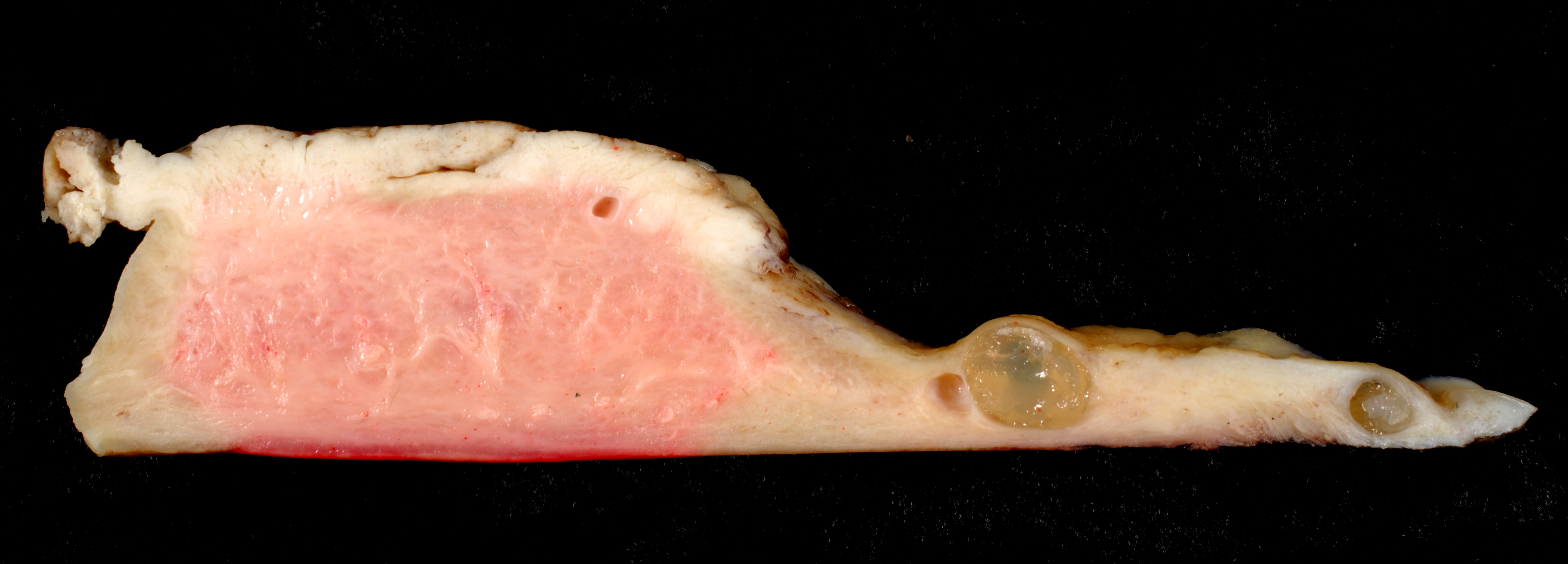
This grade-I adenocarcinoma was diagnosed by an endometrial biopsy done in the gynecologist's office. This is from the hysterectomy specimen. The tumor is limited ot the corpus uteri and does not grossly invade the underlying myometrium. This is a longitudinal sagittal section through the full length of the uterus. The corpus is to the left, and the rather elongate cervix trails off to the right. The hysterectomy specimen was bivalved, pinned out on a big bloc of paraffin wax, and floated in a vat of formalin overnight. The next morning, I cut this central section from the pinned-out half of the uterus. Top left: The carcinoma is limited to the surface of the endometrial cavity of the corpus uteri. Bottom left: No tumor is seen in the underlying myometrium. The prognosis of such non-invasive adenocarcinomas is excellent. Middle-right: Nabothian cysts are very commonly seen in the cervix and are of no clinical significance. Right: The cervix of this patient is unusually long and skinny. |
| Date |
|
| Source | https://www.flickr.com/photos/euthman/398750764/ |
| Author | Ed Uthman, MD |
| Permission (Reusing this file) |
CC-BY-SA 2.0 |
Licensing
- You are free:
- to share – to copy, distribute and transmit the work
- to remix – to adapt the work
- Under the following conditions:
- attribution – You must give appropriate credit, provide a link to the license, and indicate if changes were made. You may do so in any reasonable manner, but not in any way that suggests the licensor endorses you or your use.
- share alike – If you remix, transform, or build upon the material, you must distribute your contributions under the same or compatible license as the original.
| This image was originally posted to Flickr by euthman at https://www.flickr.com/photos/78147607@N00/398750764. It was reviewed on 1 April 2008 by FlickreviewR and was confirmed to be licensed under the terms of the cc-by-sa-2.0. |
File history
Click on a date/time to view the file as it appeared at that time.
| Date/Time | Thumbnail | Dimensions | User | Comment | |
|---|---|---|---|---|---|
| current | 17:19, 22 February 2007 | 4,205 × 1,512 (1.17 MB) | commons>Patho | == Summary == {{Information |Description=This grade-I adenocarcinoma was diagnosed by an endometrial biopsy done in the gynecologist's office. This is from the hysterectomy specimen. The tumor is limited ot the corpus uteri and does not grossly invade the |
File usage
The following page uses this file: