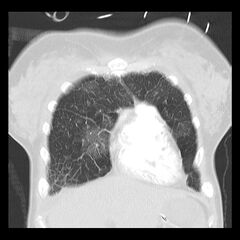
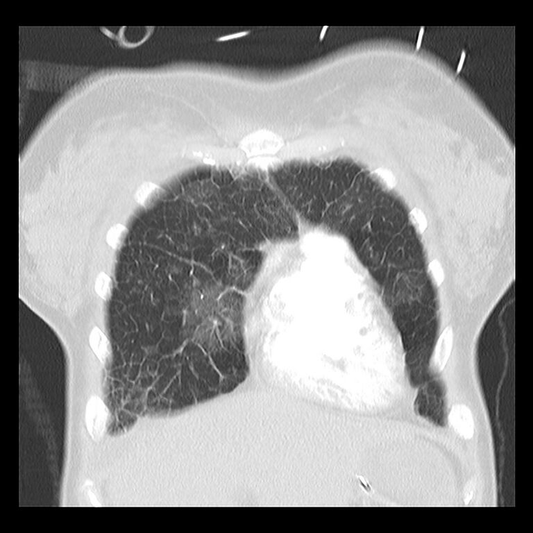
File:Acute pulmonary edema on CT (Radiopaedia 33582-34672 Coronal lung window 6).jpg

From WikiProjectMed
Jump to navigation Jump to search

Original file(1,526 × 1,526 pixels, file size: 381 KB, MIME type: image/jpeg)

This file is from a shared repository and may be used by other projects. The description on its file description page there is shown below.

Summary:

Description
  • Radiopaedia case ID: 33582
  • Image ID: 10449536
  • Image stack position: 6/36
  • Plane projection: Coronal
  • Aux modality: lung window
  • Study description: CT Pulmonary Angiogram
  • Study findings: Airspace opacity in a central peribronchovascular distribution classic of acute pulmonary edema. There is also smooth thickening of the interlobular septae in the lung bases and apices consistent with interstitial pulmonary edema and correlating with the radiographic finding of Kerley lines. There is dilatation of the main pulmonary artery and right heart enlargement / strain with straightening of the interventricular septum. Pulmonary hypertension was subsequently confirmed.
  • Modality: CT
  • System: Cardiac
  • Findings: Bilateral perihilar (batwing) airspace opacity typical of acute pulmonary edema. Kerley B lines are also well seen consistent with interstitial edema. The main pulmonary artery is dilated. ETT and NGT are appropriately positioned.
Date Published: 16th Jan 2015
Source https://radiopaedia.org/cases/acute-pulmonary-oedema-on-ct
Author Andrew Dixon
Permission
(Permission-reusing-text)
http://creativecommons.org/licenses/by-nc-sa/3.0/

Licensing:

Attribution-NonCommercial-ShareAlike 3.0 Unported (CC BY-NC-SA 3.0)

File history

Click on a date/time to view the file as it appeared at that time.

Date/TimeThumbnailDimensionsUserComment
current12:17, 18 April 2021Thumbnail for version as of 12:17, 18 April 20211,526 × 1,526 (381 KB)Radiopaedia project rID:33582 (batch #1061-6 A6)

The following page uses this file: