File:8531814250 94ef59e47c o (1).jpg

From WikiProjectMed
Jump to navigation Jump to search

8531814250_94ef59e47c_o_(1).jpg(219 × 197 pixels, file size: 13 KB, MIME type: image/jpeg)

This file is from a shared repository and may be used by other projects. The description on its file description page there is shown below.

Summary

Description
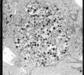
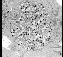
English: Left to right: MuLV found in pericyte near capillary of mouse neuronal tissue; Chlamydia pneumoniae in HeLa cell 72 hours post infection; Borrelia burgdorferi; Yersinia pestis in flea midgut.
Date
Source https://www.flickr.com/photos/niaid/8531814250/
Author Left to right: MuLV found in pericyte near capillary of mouse neuronal tissue; Chlamydia pneumoniae in HeLa cell 72 hours post infection; Borrelia burgdorferi; Yersinia pestis in flea midgut. Credit: NIAID

Licensing

{{subst:Custom license marker added by UW}} https://creativecommons.org/licenses/by/2.0/ Attribution 2.0 Generic (CC BY 2.0)

This file was uploaded with UploadWizard.

File history

Click on a date/time to view the file as it appeared at that time.

Date/TimeThumbnailDimensionsUserComment
current17:48, 3 April 2023Thumbnail for version as of 17:48, 3 April 2023219 × 197 (13 KB)Ozzie10aaaaUploaded a work by Left to right: MuLV found in pericyte near capillary of mouse neuronal tissue; Chlamydia pneumoniae in HeLa cell 72 hours post infection; Borrelia burgdorferi; Yersinia pestis in flea midgut. Credit: NIAID from https://www.flickr.com/photos/niaid/8531814250/ with UploadWizard

The following page uses this file: