File:41467 2022 28428 Fig1 HTML (1) (1).png

From WikiProjectMed
Jump to navigation Jump to search

Original file(1,123 × 496 pixels, file size: 113 KB, MIME type: image/png)

This file is from a shared repository and may be used by other projects. The description on its file description page there is shown below.

Summary

Description
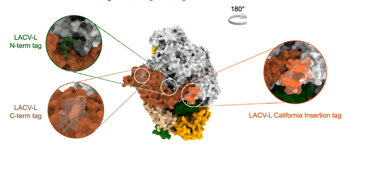
English⧼Colon⧽ English⧼Colon⧽ a Histogram of LACV-L mini-replicon activity. The percentage of Renilla/Luciferase activity of the constructs containing the N-terminal hexa-histidine tag (N-term tag), the C-terminal hexa-histidine tag (C-term tag), and the California insertion tag (Cl tag) are compared to LACV-L activity of the construct without tag (WT). Tag positions are shown on LACV-L (PDB: 6Z6G) as dotted lines. The polymerase core is colored in light gray with the Cl in orange, the cap-binding domain (CBD) in gold, the endonuclease (ENDO) in green, the mid domain in beige and the zinc-binding domain (ZBD) in brown. n = 3 biologically independent experiments. Error bars are mean ± standard deviation. Source data are provided as a Source Data file. b In vitro replication activity of LACV-LCItag_H34K using different combinations of 5′- and 3′-vRNAs. The assays are done in presence of either the 4 NTPs (AUGC), 3 NTPs (AUG) or 3 NTPs during 4 h, subsequently supplemented with CTP for 30 min (AUG+C). 5′-vRNA containing nucleotides 1 to 17 (5′ vRNA 1-17) or 5′-vRNA with base pair mutation (5′ 1-17BPm) is used. 3′-vRNA contain either nucleotides 1 to 25 (3′ vRNA 1-25) or 1 to 30 (3′ vRNA 1–30). Products have either the expected size or are extended in 5′ with GU or GGU (GU/GGU 5′ elongated products). Replication products and their respective lengths are displayed on the right side of the gel. The decade molecular weight marker (MW) is shown. Source data are provided as a Source Data file. This experiment was repeated independently 3 times with similar results. c Sequence and secondary structures of the RNA bound to LACV-LCItag_H34K for each replication state. 5′-vRNAs, 3′-vRNAs and replication products are respectively colored in pink/gray, gold and cyan. Bases present in the sequences but not seen in the structures are shown in transparent. d Cartoon representation of each LACV-LCItag_H34K replication structure. LACV-L domains are colored as in a. RNAs displayed as surfaces are colored as in a, c. The pre-initiation state corresponds to the PDB 6Z6G.
Date
Source https://www.nature.com/articles/s41467-022-28428-z
Author Benoît Arragain, Quentin Durieux Trouilleton, Florence Baudin, Jan Provaznik, Nayara Azevedo, Stephen Cusack, Guy Schoehn & Hélène Malet

Licensing

{{subst:Custom license marker added by UW}} https://creativecommons.org/licenses/by/4.0/ Attribution 4.0 International (CC BY 4.0)

This file was uploaded with UploadWizard.

File history

Click on a date/time to view the file as it appeared at that time.

Date/TimeThumbnailDimensionsUserComment
current18:58, 2 April 2023Thumbnail for version as of 18:58, 2 April 20231,123 × 496 (113 KB)Ozzie10aaaaUploaded a work by Benoît Arragain, Quentin Durieux Trouilleton, Florence Baudin, Jan Provaznik, Nayara Azevedo, Stephen Cusack, Guy Schoehn & Hélène Malet from https://www.nature.com/articles/s41467-022-28428-z with UploadWizard

There are no pages that use this file.

Metadata