File:1-s2.0-S1083879120303657-gr2.jpg

From WikiProjectMed
Jump to navigation Jump to search

1-s2.0-S1083879120303657-gr2.jpg(696 × 411 pixels, file size: 63 KB, MIME type: image/jpeg)

This file is from a shared repository and may be used by other projects. The description on its file description page there is shown below.

Summary

Description
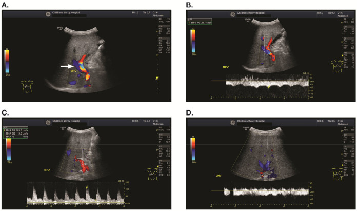
English: Figure 2. A 2-year-old male who developed VOD/SOS after HCT for acute myelogenous leukemia. (A) Color Doppler ultrasound images from an examination performed 16 days after HCT showing flow in the main portal vein away from the liver parenchyma (arrow). (B) Spectral Doppler image from the same patient showing flow away from the liver, with a peak velocity of 20.7 cm/s. (C) Spectral Doppler image from the same patient showing elevated peak systolic velocity in the proper hepatic artery causing an elevated resistive index of .83. (D) Spectral Doppler image from the same patient showing relatively constant flow in the left hepatic vein with lack of normal respiratory and cardiac variation. VOD/SOS, veno-occlusive disease/sinusoidal obstruction syndrome; HCT, hematopoietic cell transplantation.
Date
Source https://www.sciencedirect.com/science/article/pii/S1083879120303657#bib0028
Author Sherwin S. Chan , Antonio Colecchia , Rafael F. Duarte , Francesca Bonifazi , Federico Ravaioli , Jean Henri Bourhis

Licensing

English: This file is licensed CC BY-NC-ND 4.0

This file was uploaded with UploadWizard.

File history

Click on a date/time to view the file as it appeared at that time.

Date/TimeThumbnailDimensionsUserComment
current22:05, 4 November 2023Thumbnail for version as of 22:05, 4 November 2023696 × 411 (63 KB)Ozzie10aaaaUploaded a work by Sherwin S. Chan , Antonio Colecchia , Rafael F. Duarte , Francesca Bonifazi , Federico Ravaioli , Jean Henri Bourhis from https://www.sciencedirect.com/science/article/pii/S1083879120303657#bib0028 with UploadWizard

There are no pages that use this file.