Tinea nigra
| Tinea nigra | |
|---|---|
| Other names: Superficial phaeohyphomycosis, tinea nigra plantaris[1] | |
 | |
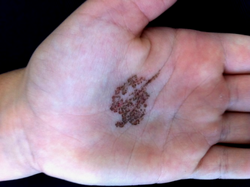
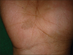
| Tinea nigra of the palm | |
| Specialty | Dermatology |
| Symptoms | One or more dark brown, painless spots on palms or soles[2] |
| Causes | Hortaea wernecki[2] |
| Diagnostic method | Visualisation, dermoscopy, microscopy, culture[1] |
| Differential diagnosis | Addison's disease, syphilis, pinta, yaws, melanoma, lentigines, palmer lichen planus, junctional melanocytes nevus[1] |
| Treatment | Antifungals, scraping the lesion[2] |
| Medication | Topical Whitfield's ointment or salicylic acid, itraconazole by mouth[1] |
| Frequency | Uncommon, female > male, children and young adults[1] |
Tinea nigra is a fungal infection of the top layer of skin.[1] It typically presents as a single 1 to 5 cm dark brown or black, non-scaly, flat, painless patch on the palms of the hand or soles of the foot.[1] There is little or no skin inflammation.[1] Typically it occurs in people who are otherwise healthy.[1]
It is a type of phaeohyphomycosis rather than a tinea.[1] Most cases are caused by Hortaea wernecki, a pigmented fungus, which is a dark yeast found in sewage, soil, rotting plant matter, and places with a high salt content such as moldy salted fish or on beaches, where contact with sand may result in transmission.[1][2] Infection is by direct contact.[1] The fungus enters and remains in the outer dead layer of skin, with no invasion of deeper tissues.[1]
Diagnosis is looking at the rash, dermoscopy, microscopy or culture of skin scrapings.[1] Other conditions that may present similarly includes Addison's disease, syphilis, pinta, yaws, melanoma, lentigines, palmer lichen planus. and junctional melanocytes nevus.[1] Treatment is with topical Whitfield's ointment or salicylic acid ointment.[1] Topical antifungals or itraconazole by mouth are other options.[3] Scraping the lesion can be curative.[2] Prevention is by general hygiene measures.[1]
Tinea nigra is uncommon.[1] It generally occurs in tropical and subtropical countries of Central or South America, the Caribbean, Europe, South East Asia, Australia, and the Far East.[1] It is rare in Japan though not uncommon in the US.[1] The disease was first described by Alexandre Cerqueira from Brazil in 1891.[1] No cases in animals have been reported.[1]
Signs and symptoms
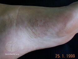
It typically presents as a single 1 to 5cm dark brown-black, non-scaly, flat, painless patch on the palms of the hands and the soles of the feet of healthy people.[1] There may be multiple spots.[1]
-

-
-
-
Brown colored macule of tinea nigra


.jpg)

Causes
Tinea nigra is caused by the fungus formerly classified as Cladosporium werneckii, but more recently classified as Hortaea werneckii.[4] The causative organism has also been described as Phaeoannellomyces werneckii.[5] Tinea nigra is extremely superficial and can be removed from the skin by forceful scraping. It tends to appear in areas where eccrine sweat glands are highly concentrated. Infections generally start to appear on the skin around 2–7 weeks post inoculation.
The ability of H. werneckii to tolerate high salt concentrations and acidic conditions allows it to flourish inside the stratum corneum. H. wernickii tends remain localized in one spot or region, and produces darkly-colored, brown macules on the skin due to the production of a melanin-like substance.[6]
Diagnosis

Diagnosis of tinea nigra is made based on microscopic examination of stratum corneum skin scrapings obtained by using a scalpel. The scrapings are mixed with potassium hydroxide (KOH).[7] The KOH lyses the nonfungal debris.[7] The skin scrapings are cultured on Sabouraud's agar at 25°C and allowed to grow for about a week. H. werneckii can generally be distinguished due to its two-celled yeast form and the presence of septate hyphae with thick, darkly pigmented walls.[6]
Treatment
Treatment consists of topical application of dandruff shampoo, which contains selenium sulfide, over the skin. Topical antifungal imidazoles such as ketoconazole, itraconazole, and miconazole may also be used. Imidazoles are generally used twice daily for a two-week period. This is the same treatment plan for tinea or pityriasis versicolor. Other treatment methods include the use of epidermal tape stripping, Undecylenic acid, and other topical agents such as ciclopirox. Once a tinea nigra infection has been eradicated from the host, it is not likely to reoccur.[6]
Epidemiology
Tinea nigra is commonly found in Africa, Asia, Central America, and South America. It is typically not found in the United States or Europe, although cases have been documented in the Southeastern United States. People of all ages can be infected; however, it is generally more apparent in children and younger adults. Females are three times more likely than males to become infected.[6]
See also
References
- ↑ 1.00 1.01 1.02 1.03 1.04 1.05 1.06 1.07 1.08 1.09 1.10 1.11 1.12 1.13 1.14 1.15 1.16 1.17 1.18 1.19 1.20 1.21 1.22 1.23 Chander, Jagdish (2018). "8. Tinea Nigra". Textbook of Medical Mycology (4th ed.). New Delhi: Jaypee Brothers Medical Publishers Ltd. pp. 145–153. ISBN 978-93-86261-83-0. Archived from the original on 2021-10-02. Retrieved 2021-10-02.
- ↑ 2.0 2.1 2.2 2.3 2.4 James, William D.; Elston, Dirk; Treat, James R.; Rosenbach, Misha A.; Neuhaus, Isaac (2020). "15. Diseases resulting from fungi and yeasts". Andrews' Diseases of the Skin: Clinical Dermatology (13th ed.). Elsevier. p. 299. ISBN 978-0-323-54753-6. Archived from the original on 2023-03-23. Retrieved 2023-03-23.
- ↑ Morris-Jones, Rachael (2019). "18. Tropical dermatology". In Morris-Jones, Rachael (ed.). ABC of Dermatology (7th ed.). Hoboken: Wiley Blackwell. p. 151. ISBN 978-1-119-48899-6. Archived from the original on 2022-05-16. Retrieved 2022-05-17.
- ↑ Murray, Patrick R.; Rosenthal, Ken S.; Pfaller, Michael A. (2005). Medical Microbiology (5th ed.). Elsevier Mosby.
- ↑ Pegas JR, Criado PR, Lucena SK, de Oliveira MA (2003). "Tinea nigra: report of two cases in infants". Pediatric Dermatology. 20 (4): 315–7. doi:10.1046/j.1525-1470.2003.20408.x. PMID 12869152.
- ↑ 6.0 6.1 6.2 6.3 Schwartz, Robert A (September 2004). "Superficial fungal infections". The Lancet. 364 (9440). doi:10.1016/S0140-6736(04)17107-9. PMID 15451228.
- ↑ 7.0 7.1 Gladwin, Mark; Trattler, Bill. Clinical Microbiology (4th ed.). p. 196.
External links
| Classification | |
|---|---|
| External resources |