Supraorbital vein
| Supraorbital vein | |
|---|---|
 Veins of the head and neck. (Supraorbital vein visible at upper right.) | |
 The tarsi and their ligaments. Right eye; front view. (Supraorbital vessels labeled at upper right.) | |
| Details | |
| Drains from | forehead |
| Drains to | angular |
| Artery | supra-orbital |
| Identifiers | |
| Latin | vena supraorbitalis |
| TA98 | A12.3.05.021 |
| TA2 | 4820 |
| FMA | 50904 |
| Anatomical terminology | |
The supraorbital vein is a vein of the forehead. It communicates with the frontal branch of the superficial temporal vein. It passes through the supraorbital notch, and merges with the angular vein to form the superior ophthalmic vein. The supraorbital vein helps to drain blood from the forehead, eyebrow, and upper eyelid.
Structure
The supraorbital vein begins on the forehead, where it communicates with the frontal branch of the superficial temporal vein. It runs downward superficial to the frontalis muscle. It merges with the angular vein to form the superior ophthalmic vein.[1][2]
Previous to its junction with the angular vein, it passes through the supraorbital notch into the orbit around the eye.[1] As this vessel passes through the notch, it receives the frontal diploic vein through a foramen at the bottom of the notch.
Function
The supraorbital vein helps to drain blood from the forehead, eyebrow, and upper eyelid.
Additional images
-
 Bloodvessels of the eyelids, front view.
 -
 Lateral head anatomy detail
 -
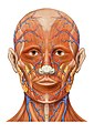 Head anatomy anterior view


References
![]() This article incorporates text in the public domain from page 645 of the 20th edition of Gray's Anatomy (1918)
This article incorporates text in the public domain from page 645 of the 20th edition of Gray's Anatomy (1918)
- ^ a b Remington, Lee Ann (2012). "11 - Orbital Blood Supply". Clinical Anatomy and Physiology of the Visual System (3rd ed.). Butterworth-Heinemann. pp. 202–217. doi:10.1016/B978-1-4377-1926-0.10011-6. ISBN 978-1-4377-1926-0.
- ^ Dutton, Jonathan J. (2010). "3 - Anatomy of the Orbit". Radiology of the Orbit and Visual Pathways. Saunders. pp. 31–39. doi:10.1016/B978-1-4377-1151-6.00003-3. ISBN 978-1-4377-1151-6.
This cardiovascular system article is a stub. You can help Wikipedia by expanding it. |
- Articles with short description
- Short description matches Wikidata
- Anatomy NAV infobox with use of other NAV parameters
- Wikipedia articles incorporating text from the 20th edition of Gray's Anatomy (1918)
- Articles with TA98 identifiers
- All stub articles
- Cardiovascular system stubs
- Veins of the head and neck