Spider angioma
| Spider angioma | |
|---|---|
| Other names: Nevus araneus, spider nevus, vascular spider, spider telangiectasia[1] | |
 | |
| Non-benign angiomas indicating cirrhosis | |
| Specialty | Dermatology |
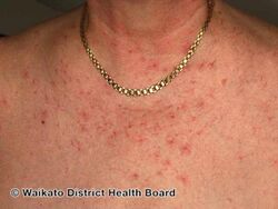
A spider angioma or spider naevus (plural: spider naevi), also nevus araneus, is a type of telangiectasis[2] (swollen blood vessels) found slightly beneath the skin surface, often containing a central red spot and deep reddish extensions (see Blood color) which radiate outwards like a spider's web or a spider's legs. They are common and often benign, presenting in around 10–15% of healthy adults and young children.[3] However, having more than three spider angiomas is likely to be abnormal and may be a sign of liver disease; it also suggests the probability of esophageal varices.[4]
Signs and symptoms
Spider angiomas are found only in the distribution of the superior vena cava, and are thus commonly found on the face, neck, upper part of the torso, and arms.
-
Spider naevi
-
![Nevus araneus (spider nevus): in the center of the red lesion a small (1 mm) red papule is visible, surrounded by several distinct radiating vessels. Pressure on the lesion causes it to disappear, however blanching is replaced by rapid refill from the central arteriole when pressure is released.[5]](https://upload.wikimedia.org/wikipedia/commons/thumb/6/68/Spidernevus.jpg/250px-Spidernevus.jpg)
-

Multiple spider angiomas
.jpg)
![Nevus araneus (spider nevus): in the center of the red lesion a small (1 mm) red papule is visible, surrounded by several distinct radiating vessels. Pressure on the lesion causes it to disappear, however blanching is replaced by rapid refill from the central arteriole when pressure is released.[5]](/wiki/File:Spidernevus.jpg)

Cause
Spider angiomas form due to failure of the sphincteric muscle surrounding a cutaneous arteriole. The central red dot is the dilated arteriole and the red "spider legs" are small capillaries carrying away the freely flowing blood. If momentary pressure is applied, it is possible to see the emptied capillaries refilling from the center. No other angiomas show this phenomenon.[6]
The dilation, in turn, is caused by increased estrogen levels in the blood. Many pregnant women and women using hormonal contraception have spider angiomas, which is due to high estrogen levels in their blood. Individuals with significant liver disease also show many spider angiomas, as their liver cannot metabolize circulating estrogens, specifically estrone, which derives from the androgen androstenedione.[3] About 33% of patients with cirrhosis have spider angiomas.[7]
Diagnosis
Diagnosis is by clinical examination. Spider naevi are most commonly seen by general practitioners, or dermatologists. Whilst a lesion can be identified as a spider naevus, this is not a diagnosis in itself. The clinical picture should be indicative of whether there is underlying disease that should be investigated.[citation needed]
Treatment
Spider angiomas are asymptomatic and usually resolve spontaneously. This is common in the case of children, although they may take several years to disappear. If the spider angiomas are associated with pregnancy, they may resolve after childbirth. In women taking oral contraceptives, they may resolve after stopping these contraceptives.
For spider angiomas on the face, techniques such as electrodesiccation and laser treatment can be used to remove the lesion.[8] There is a small risk of a scar, however it usually leaves nothing.
See also
References
- ↑ Rapini, Ronald P.; Bolognia, Jean L.; Jorizzo, Joseph L. (2007). Dermatology: 2-Volume Set. St. Louis: Mosby. pp. 1621–22. ISBN 978-1-4160-2999-1.
- ↑ "spider angioma" at Dorland's Medical Dictionary
- ↑ 3.0 3.1 Nevus Araneus at eMedicine
- ↑ Udell, Jacob A.; Wang, Charlie S.; Tinmouth, Jill; FitzGerald, J. Mark; Ayas, Najib T.; Simel, David L.; Schulzer, Michael; Mak, Edwin; Yoshida, Eric M. (22 February 2012). "Does This Patient With Liver Disease Have Cirrhosis?". JAMA. 307 (8): 832–842. doi:10.1001/jama.2012.186. PMID 22357834. Retrieved 11 April 2018 – via jama.jamanetwork.com.
- ↑ Sand, M; Sand, D; Thrandorf, C; Paech, V; Altmeyer, P; Bechara, FG (4 June 2010). "Cutaneous lesions of the nose". Head & Face Medicine. 6: 7. doi:10.1186/1746-160X-6-7. PMC 2903548. PMID 20525327.
- ↑ McCluskey D R Journal of the Royal College of Physicians of Edinburgh 2004, 34: 104 - 105
- ↑ Li CP, Lee FY, Hwang SJ, et al. (1999). "Spider angiomas in patients with liver cirrhosis: role of alcoholism and impaired liver function". Scand. J. Gastroenterol. 34 (5): 520–3. doi:10.1080/003655299750026272. PMID 10423070.
- ↑ Geronemus, R. G. (1991). "Treatment of spider telangiectases in children using the flashlamp-pumped pulsed dye laser". Pediatr Dermatol. 8 (1): 61–3. doi:10.1111/j.1525-1470.1991.tb00843.x. PMID 1862028.
External links
| Classification | |
|---|---|
| External resources |