Right-sided aortic arch
| Right-sided aortic arch | |
|---|---|
 | |
| Anterior-posterior chest radiograph showing a right-sided aortic arch | |
Right-sided aortic arch is a rare anatomical variant in which the aortic arch is on the right side rather than on the left. During normal embryonic development, the aortic arch is formed by the left fourth aortic arch and the left dorsal aorta. In people with a right-sided aortic arch, instead the right dorsal aorta persists and the distal left aorta disappears.[citation needed]
Symptoms and signs
A right-sided aortic arch does not cause symptoms on itself, and the overwhelming majority of people with the right-sided arch have no other symptoms. However when it is accompanied by other vascular abnormalities, it may form a vascular ring, causing symptoms due to compression of the trachea and/or esophagus.[1]
Pathophysiology
The causes of right-sided aortic arch are still unknown, 22q11 deletions have been found in some people with this condition.[2] It has also been found in association with other genetic syndromes such as Trisomy 21 (Down syndrome).[citation needed]
Diagnosis
-
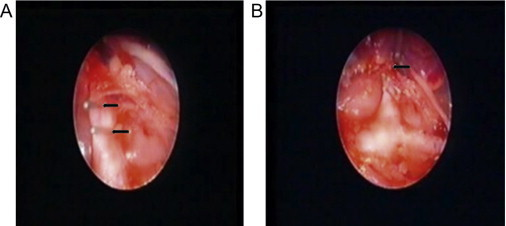
a) Right-sided aortic arch with abberant left subclavian artery – clipped b) right-sided aortic arch with abberant left subclavian artery – divided
-
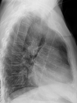
Chest radiograph showing a right-sided aortic arch, lateral view


During pregnancy, prenatal ultrasound may reveal the abnormal course of the arch and this is the most common reason for identification of a right sided aortic arch nowadays.[3] Sometimes, when a right sided aortic arch is seen before birth, it can actually be a double aortic arch, sometimes a fetal MRI scan may be helpful if the ultrasound is not clear.[4]
After birth, a right-sided aortic arch is visualized on chest radiography, by the aortic knob (the prominent shadow of the aortic arch) that is located right from the sternum instead of left. Complex lesions are often assessed by MRI or CT.[citation needed]
Classification
Several types of right-sided aortic arch exist, the most common ones being right-sided aortic arch with aberrant left subclavian artery and the mirror-image type. The variant with aberrant left subclavian artery is associated with congenital heart disease in only a small minority of affected people. The mirror-image type of right aortic arch is very strongly associated with congenital heart disease, in most cases tetralogy of Fallot.[citation needed]
Management
If a right aortic arch is associated with a left sided arterial ductal ligament (a remnant from the foetal circulation which forms a ligament after birth) then a vascular ring is formed around the trachea. Studies show that around 1:4 children show symptoms of a vascular ring.[5] This requires further investigation by specialists. Many children are well. There is evidence to show that symptoms of a vascular ring do not correlate with the appearance of the trachea in these patients so further assessment may be required. [6]This could be in the form of a specialist CT scan which is timed with inspiration and expiration or a free breathing bronchoscopy.[citation needed]
If required, repairing a vascular ring formed by a right sided aortic arch usually involves dividing the left sided arterial ductal ligament (this is not a structure that is necessary for the heart circulation as it is not a vessel after birth). This is usually performed by cardiothoracic surgeons from the side of the chest (thoracotomy incision) and does not require the heart to be stopped like many heart surgeries. Some people may have an aberrant left subclavian artery (the artery to the left arm) and this may also require re-implantation as it adds to the complexity of the vascular ring.[citation needed]
Epidemiology
Right-sided aortic arch is rare, with a prevalence among adults of about 0.01%.[citation needed]
See also
References
- ↑ Białowas J, Hreczecha J, Grzybiak M (2000). "Right-sided aortic arch". Folia Morphol. (Warsz). 59 (3): 211–6. PMID 10974792.
- ↑ Momma, K.; Matsuoka, R.; Takao, A. (2014). "Aortic Arch Anomalies Associated with Chromosome 22q11 Deletion (CATCH 22)". Pediatric Cardiology. 20 (2): 97–102. doi:10.1007/s002469900414. ISSN 0172-0643. PMID 9986884. S2CID 813344.
- ↑ Zidere V, Tsapakis EG, Huggon IC, Allan LD (December 2006). "Right aortic arch in the fetus". Ultrasound Obstet Gynecol. 28 (7): 876–81. doi:10.1002/uog.3841. PMID 17066500. S2CID 25566821.
- ↑ "Spotlight on research | News Centre | King's College London". Archived from the original on 2018-07-31. Retrieved 2022-03-04.
- ↑ Fetuses with right aortic arch: a multicenter cohort study and meta-analysis. D'Antonio F, Khalil A, Zidere V, Carvalho JS. Ultrasound Obstet Gynecol. 2016 Apr;47(4):423-32. doi: 10.1002/uog.15805. Epub 2016 Mar 16. Review.
- ↑ Correlation of Symptoms with Bronchoscopic Findings in Children with a Prenatal Diagnosis of a Right Aortic Arch and Left Arterial Duct. Vigneswaran TV, Kapravelou E, Bell AJ, Nyman A, Pushparajah K, Simpson JM, Durward A, Zidere V. Pediatr Cardiol. 2018 Apr;39(4):665-673. doi: 10.1007/s00246-017-1804-5. Epub 2018 Jan 6.
External links
- Radiopaedia: Aortic arch variants Archived 2013-02-19 at the Wayback Machine
- eMedicine: Right Aortic Arch in Vascular Ring Defects Archived 2021-07-26 at the Wayback Machine
- Pages with script errors
- All articles with unsourced statements
- Articles with unsourced statements from October 2021
- Articles with invalid date parameter in template
- Articles with unsourced statements from January 2021
- Articles with unsourced statements from February 2021
- Webarchive template wayback links
- Congenital vascular defects