Rhinosporidiosis
| Rhinosporidiosis | |
|---|---|
 | |
| Rhinosporidiosis in oropharynx | |
| Specialty | Infectious disease |
Rhinosporidiosis is an infection caused by Rhinosporidium seeberi.[1][2]
Classification
This organism was previously considered to be a fungus, and rhinosporidiosis is classified as a fungal disease under ICD-10. It is now considered to be a protist[3] classified under Mesomycetozoea.[4]
Authors of detailed studies have revealed superficial similarities between Dermocystidium and Rhinosporidium when using light microscopy, but substantial morphological differences between the groups exist.[5]
There is some evidence that DNA extracted from purified uncontaminated round bodies (Rhinosporidium seeberi) is of cyanobacterial origin.[6]
Signs and symptoms
The clinical presentation of Rhinosporidiosis is as follows:[7]
- Itching
- Photophobia
- Conjunctivitis
- Bloody tears
Pathophysiology
Rhinosporidiosis is a granulomatous disease affecting the mucous membrane of nasopharynx, oropharynx, conjunctiva, rectum and external genitalia. Though the floor of the nose and inferior turbinate are the most common sites, the lesions may appear elsewhere too. Traumatic inoculation from one site to others is common. Laryngeal rhinosporidiosis,[8] too, has been described and may be due to inoculation from the nose during endotracheal intubation. After inoculation, the organism replicates locally, resulting in hyperplasia of host tissue and localised immune response.
- infection of nose and nasopharynx - 70%
- infection of palpebral conjunctiva - 15%
Diagnosis
-
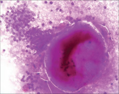
Cytology smear shows a ruptured sporangium with many endospores
-
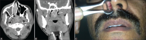
Contrast-enhanced CT PNS a) Axial image shows an enhancing soft tissue lesion (black arrow) in the right inferior nasal cavity extending through the choana into the nasopharynx. b) Coronal section shows lobulated nasopharyngeal extension of the lesion (white arrow) with prominent leash of blood vessels. c) Anterior rhinoscopy shows a red fleshy mass with whitish spots in the right nasal cavity. Diagnosis after surgery confirmed as rhinosporidiosis


The diagnosis of for this condition is done via the following:[citation needed]
- History
- Unilateral nasal obstruction
- Epistaxis
- Local pruritus
- Rhinorrhea
- Coryza (rhinitis) with sneezing
- Post nasal discharge with cough
- Foreign body sensation
- History of exposure to contaminated water
- Increased tearing and photo phobia in cases of infection of palpebral conjunctiva
- On examination
- Pink to deep red polyps
- Strawberry like appearance
- Bleeds easily upon manipulation
- Diagnosis
- confirmed by biopsy and histopathology - several round or oval sporangia and spores which may be seen bursting through its chitinous wall
Treatment
- Surgical excision - wide excision with wide area electro-coagulation of the lesion base
- Medical treatment is not so effective but treatment with a year-long course of dapsone has been reported
- Recurrence is common
Epidemiology
Disease endemic in Chhattisgarh South India, Sri Lanka, South America and Africa. It is presumed to be transmitted by exposure to the pathogen when taking a bath in stagnant water pools where animals also bathe.
References
- ↑ Arseculeratne SN (2002). "Recent advances in rhinosporidiosis and rhinosporidium seeberi". Indian J Med Microbiol. 20 (3): 119–31. PMID 17657050. Archived from the original on 2021-01-27. Retrieved 2021-06-17.
- ↑ Arseculeratne SN (April 2005). "Rhinosporidiosis: what is the cause?". Curr. Opin. Infect. Dis. 18 (2): 113–8. doi:10.1097/01.qco.0000160898.82115.e8. PMID 15735413.
- ↑ Morelli L, Polce M, Piscioli F, et al. (2006). "Human nasal rhinosporidiosis: an Italian case report". Diagn Pathol. 1 (1): 25. doi:10.1186/1746-1596-1-25. PMC 1560165. PMID 16945122.
- ↑ "Rhinosporidiosis". Archived from the original on 2021-09-23. Retrieved 2021-06-17.
- ↑ Pekkarinen, Low, Murphy, Ragan and Dykova. 2003. Phylogenetic position and ultrastructure of two Dermocystidium species are(Ichthyosporea) from the common perch (Perca fluviatilis) Archived 2007-10-10 at the Wayback Machine. Acta Protozoologica Vol. 42:287-307
- ↑ Dhaulakhandi, Ahluwalia, Ravi and Garg. 2006. Detection of 16S rRNA gene in round bodies isolated from polyps of rhinosporidiosis. Infection, Genetics and Evolution Vol. 6:331-336
- ↑ "Rhinosporidiosis - EyeWiki". eyewiki.aao.org. Archived from the original on 27 May 2022. Retrieved 4 November 2022.
- ↑ Ajit Daharwal, Hansa Banjara, Digvijay Singh, Anuj Gupta, Surjeet Singh. 2011. A rare case of laryngeal rhinosporidiosis Archived 2021-03-09 at the Wayback Machine. J Laryngol Voice 2011;1:30-2
External links
| Classification | |
|---|---|
| External resources |