Pott disease
| Pott disease | |
|---|---|
 | |
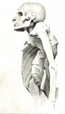
| Tuberculosis of the spine in an Egyptian mummy | |
| Symptoms | Pott's spine, tuberculous spondylitis, spinal tuberculosis |
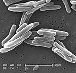
Pott disease is tuberculosis of the spine,[1] usually due to haematogenous spread from other sites, often the lungs. The lower thoracic and upper lumbar vertebrae areas of the spine are most often affected.
It causes a kind of tuberculous arthritis of the intervertebral joints. The infection can spread from two adjacent vertebrae into the adjoining intervertebral disc space. If only one vertebra is affected, the disc is normal, but if two are involved, the disc, which is avascular, cannot receive nutrients, and collapses. In a process called caseous necrosis, the disc tissue dies, leading to vertebral narrowing and eventually to vertebral collapse and spinal damage. A dry soft-tissue mass often forms and superinfection is rare.
Spread of infection from the lumbar vertebrae to the psoas muscle, causing abscesses, is not uncommon.[2]
The disease is named after Percivall Pott, the British surgeon who first described it in the late 18th century.
Signs and symptoms
The clinical presentation of this condition is as follows:[3]
- Back pain
- Weight loss
- Fever
- Fatigue
Mechanism
Primary site of tuberculosis infection ( lungs) spreads via paradiscal vessels on either side of disc space. Progressive vertebral loss can cause spinal kyphotic deformity.[3]
Diagnosis
- – Complete blood count: leukocytosis
- – Elevated erythrocyte sedimentation rate: >100 mm/h
- – Tuberculin skin test (purified protein derivative [PPD]) results are positive in 84–95% of patients with Pott disease who are not infected with HIV.

- Radiographs of the spine
- – Radiographic changes associated with Pott disease present relatively late. These radiographic changes are characteristic of spinal tuberculosis on plain radiography:
- Lytic destruction of anterior portion of vertebral body
- Increased anterior wedging
- Collapse of vertebral body
- Reactive sclerosis on a progressive lytic process
- Enlarged psoas shadow with or without calcification
- – Additional radiographic findings may include:
- Vertebral end plates are osteoporotic.
- Intervertebral disks may be shrunken or destroyed.
- Vertebral bodies show variable degrees of destruction.
- Fusiform paravertebral shadows suggest abscess formation.
- Bone lesions may occur at more than one level.
- Bone scan
- Computed tomography of the spine
- Bone biopsy
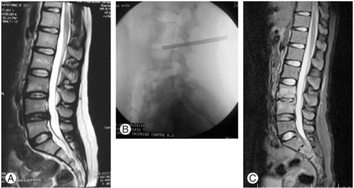
- MRI
Prevention
Controlling the spread of tuberculosis infection can prevent tuberculous spondylitis and arthritis. Patients who have a positive PPD test (but not active tuberculosis) may decrease their risk by properly taking medicines to prevent tuberculosis. To effectively treat tuberculosis, patients must take their medications exactly as prescribed.[citation needed]
Management

Treatment is based on the following:
- Nonoperative – antituberculous drugs
- Analgesics
- Immobilization of the spine region using different types of braces and collars
- Surgery may be necessary, especially to drain spinal abscesses or debride bony lesions fully or to stabilize the spine. A 2007 review found just two randomized clinical trials with at least one-year follow-up that compared chemotherapy plus surgery with chemotherapy alone for treating people diagnosed with active tuberculosis of the spine. As such, no high-quality evidence exists, but the results of this study indicates that surgery should not be recommended routinely and clinicians have to selectively judge and decide on which patients to operate.[4]
- Thoracic spinal fusion with or without instrumentation as a last resort.
- Physical therapy for pain-relieving modalities, postural education, and teaching a home-exercise program for strength and flexibility
Prognosis
- Vertebral collapse resulting in kyphosis
- Spinal cord compression
- Sinus formation
- Paraplegia (so-called Pott's paraplegia)
Culture
- In Ernest Poole's Pulitzer Prize-winning novel, His Family, young Johnny Geer has a terminal case of Pott disease.
- Passionist saint Gemma Galgani had tuberculosis of the spine.
- The fictional Hunchback of Notre Dame has a gibbus deformity similar to the type caused by tuberculosis.
- In Henrik Ibsen's play A Doll's House, Dr. Rank has "consumption of the spine".
- Jocelin, the dean who wanted a spire on his cathedral in William Golding's The Spire, probably died as a result of the disease.
- English poets Alexander Pope and William Ernest Henley both had Pott disease.
- Anna Roosevelt Cowles, sister of President Theodore Roosevelt, had Pott disease.
- Søren Kierkegaard may have died from Pott disease, according to professor Kaare Weismann and literature scientist Jens Staubrand[5]
- Chick Webb, a swing-era drummer and band leader, was afflicted with tuberculosis of the spine as a child, which left him hunchbacked, and eventually caused his death.
- The Sicilian mafia boss Luciano Leggio had the disease and wore a brace.
- Morton, the railroad magnate in Once Upon a Time in the West, has the disease and needs crutches to walk.
- Writer Max Blecher had Pott disease. His story is portrayed in the 2016 film Scarred Hearts.
- Marxist thinker and Communist leader Antonio Gramsci had Pott disease, probably due to the bad conditions of his incarceration in fascist Italy during the 1930s.
- Italian writer, poet, and philosopher Giacomo Leopardi had the disease.
- It features prominently in the book This Is a Soul, which chronicles the work of American physician Rick Hodes in Ethiopia.
- Imogen, in the novella "The Princess with the Golden Hair", part of Memoirs of Hecate County by Edmund Wilson (1946), has Pott disease.
- Jane Addams, social activist and Nobel Peace Prize winner, had Pott disease.
- Gavrilo Princip, who assassinated Archduke Franz Ferdinand of Austria, leading to World War I, died in prison of bone tuberculosis.
- Saint Bernadette of Lourdes had tuberculosis of the bone in her right knee
- In the story Two Gentlemen of Verona, written by A. J. Cronin, Lucia had tuberculosis of spine.
- Willem Ten Boom, brother of Corrie Ten Boom, died of tuberculosis of the spine in December 1946[6]
- English writer Denton Welch (1915–1948) died of spinal tuberculosis after being involved in a motor accident (1935) that irreparably damaged his spine.
- Louis Joseph, Dauphin of France, son of King Louis XVI and Marie Antoinette[7]
References
- ↑ Garg, RK; Somvanshi, DS (2011). "Spinal tuberculosis: a review". The Journal of Spinal Cord Medicine. 34 (5): 440–54. doi:10.1179/2045772311Y.0000000023. PMC 3184481. PMID 22118251.
- ↑ Wong-Taylor, LA; Scott, AJ; Burgess, H (20 May 2013). "Massive TB psoas abscess". BMJ Case Reports. 2013: bcr2013009966. doi:10.1136/bcr-2013-009966. PMC 3670072. PMID 23696148.
- ↑ 3.0 3.1 Viswanathan, Vibhu Krishnan; Subramanian, Surabhi (2021). "Pott Disease". StatPearls. StatPearls Publishing. Archived from the original on 20 January 2021. Retrieved 29 August 2021.
- ↑ Jutte PC, van Loenhout-Rooyackers JH. Routine surgery in addition to chemotherapy for treating spinal tuberculosis. Cochrane Database of Systematic Reviews 2006, Issue 1. Art. No.: CD004532. DOI: 10.1002/14651858.CD004532.pub2. http://onlinelibrary.wiley.com/doi/10.1002/14651858.CD004532.pub2/abstract Archived 2016-03-05 at the Wayback Machine
- ↑ Krasnik, Benjamin (2013). "Kierkegaard døde formentlig af Potts sygdom" (in dansk). Kristeligt Dagblad. Archived from the original on 2016-10-13. Retrieved 2016-10-02.
- ↑ The Hiding Place, Chapter: "Since Then"
- ↑ Covington, Richard. "Marie Antoinette". Smithsonian. Archived from the original on 2021-04-11. Retrieved 2019-08-18.
External links
- Pott Disease — Tuberculous Spondylitis (medical article with MRI picture), eMedicine, 2018-08-30, archived from the original on 2008-10-19, retrieved 2021-06-20.
- "Tuberculous arthritis", MedlinePlus, USA: NIH, archived from the original on 2012-02-04, retrieved 2021-06-20. Public domain.
- Pott's Disease of the Thoracic Spine Archived 2017-07-16 at the Wayback Machine
| Classification |
|---|
