Portal:Viruses/Selected picture
The following images are currently featured as the Selected picture of the Viruses portal. To suggest an image for inclusion, use the suggestions page
| Portal:Viruses/Selected picture/1
1802 cartoon of Edward Jenner administering cowpox vaccine against smallpox, satirising contemporary fears about vaccination. Credit: James Gillray (12 June 1802) |
| Portal:Viruses/Selected picture/2
Tobacco mosaic virus was the first virus to be identified, as an infectious agent that could pass through porcelain filters, as well as the first to be crystallised. It was among the earliest virus structures to be modelled successfully. Credit: Thomas Splettstoesser (20 July 2012) |
| Portal:Viruses/Selected picture/3
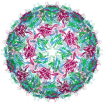
The MS2 bacteriophage was the first virus genome to be sequenced in 1976. Its capsid has an icosahedral structure made up from 180 copies of the coat protein. Credit: Neil Ranson (7 June 2011) |
| Portal:Viruses/Selected picture/4
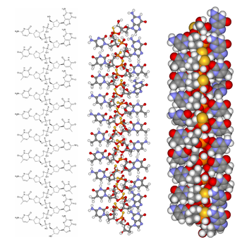
ΦX174 is a bacteriophage whose DNA genome size of 5386 nucleotides, among the smallest of DNA viruses, has led to it being the subject of pioneering research in molecular biology. Credit: Fdardel (21 March 2009) |
| Portal:Viruses/Selected picture/5
An HIV protease inhibitor (white) bound to its target, the HIV-1 protease (blue and green). The viral enzyme is a dimer of two identical subunits with the active site (red) in the cleft between them. Credit: Boghog (28 June 2008) |
| Portal:Viruses/Selected picture/6
HIV-1 budding from lymphocytes in culture. HIV establishes a latent infection in several types of immune cell and causes profound immunodeficiency. Credit: C. Goldsmith (1984) |
| Portal:Viruses/Selected picture/7
Rinderpest was a Morbillivirus that caused catastrophic cattle plagues for centuries. It was declared the second virus to have been eradicated globally in 2011. Credit: Jacobus Eussen (1745) |
| Portal:Viruses/Selected picture/8
Respiratory droplets, such as those expelled during a sneeze, are important in the transmission of several respiratory viruses, including influenza and severe acute respiratory syndrome coronavirus 2. Droplets are also released by breathing, talking, coughing and vomiting, and can be created by aerosol-generating medical procedures. Credit: James Gathany (2009) |
| Portal:Viruses/Selected picture/9
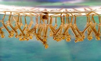
Aedes aegypti can transmit the chikungunya, dengue, yellow fever and Zika viruses. The mosquito is widespread in tropical and subtropical regions, with mosquito control being key to disease prevention. Credit: United States Department of Agriculture (2000) |
| Portal:Viruses/Selected picture/10
Ebola virus is a filamentous RNA virus first recognised in 1976. Four of the five known members of the Ebolavirus genus cause a severe haemorrhagic fever in humans. Credit: Cynthia Goldsmith |
| Portal:Viruses/Selected picture/11
The Varroa destructor mite can transmit viruses, including deformed wing virus, to its honey bee host. This might contribute to colony collapse disorder, in which worker bees abruptly disappear. Credit: Eric Erbe & Christopher Pooley |
| Portal:Viruses/Selected picture/12
Bacteriophages, viruses that infect bacteria, are among the most common entities on Earth. Credit: Graham Beards (21 October 2008) |
| Portal:Viruses/Selected picture/13
This 18th Dynasty Egyptian stele, believed to show a priest with poliomyelitis-associated deformity, is one of the earliest records of a viral disease. Credit: Unknown (1580–1350 BC) |
| Portal:Viruses/Selected picture/14
Lady Mary Wortley Montagu survived smallpox, and popularised the Turkish practice of variolation against the disease in western Europe in the 1720s. Credit: Jean-Étienne Liotard (c. 1756) |
| Portal:Viruses/Selected picture/15
Louis Pasteur invented a vaccine against rabies, and tested it on a boy bitten by a rabid dog in 1885. Credit: Albert Edelfelt (1885) |
| Portal:Viruses/Selected picture/16
Culex species mosquitoes transmit West Nile virus. Elimination of the stagnant water pools where the mosquitoes breed, together with other mosquito control measures, is key to preventing disease. Credit: James Gathany (28 February 2006) |
| Portal:Viruses/Selected picture/17
Many small icosahedral viruses have capsids made up of multiple copies of just two proteins. The proteins aggregate into units called capsomeres, which have either pentagonal or hexagonal symmetry (as shown here). Credit: Antares42 (4 September 2009) |
| Portal:Viruses/Selected picture/18
The first immortal human cell line, HeLa cells were derived from a cervical cancer biopsy and carry human papillomavirus 18 DNA. The cells have been growing since 1951. Credit: Gerry Shaw (8 March 2012) |
| Portal:Viruses/Selected picture/19
The common cold is the most frequent infectious disease. Despite the advice to "consult your physician" no antiviral treatment has been approved, and colds are only rarely associated with serious complications. Credit: Federal Art Project (1937) |
| Portal:Viruses/Selected picture/20
Some viruses, such as the T7 bacteriophage, encode their own RNA polymerase, the enzyme that makes messenger RNA based on a DNA template. The T7 enzyme has a single subunit, and is more like chloroplast and mitochondrial enzymes than those of bacteria or the cell. Credit: Thomas Splettstoesser (25 June 2007) |
| Portal:Viruses/Selected picture/21
Biosafety level 3 equipment is used for research with viruses such as influenza that can cause serious disease but for which treatment is available. The biosafety cabinet uses HEPA filters to filter viruses out of the air. This researcher is examining reconstructed 1918 pandemic influenza virus, or "Spanish flu". Credit: CDC (2005) |
| Portal:Viruses/Selected picture/22
Respiratory failure in bulbar and bulbospinal polio condemned many patients to one or two weeks in an "iron lung" or negative-pressure ventilator. The first ventilator designed for polio patients appeared in 1918; this model dates from the 1950s. Credit: Hewa (December 2011) |
| Portal:Viruses/Selected picture/23
The antiviral fomivirsen was the first antisense therapy to be licensed by the FDA. It binds to a cytomegalovirus mRNA and is used to treat cytomegalovirus retinitis. Credit: Fvasconcellos (1 January 2007) |
| Portal:Viruses/Selected picture/24
Chikungunya virus is an alphavirus transmitted by Aedes mosquitoes. The disease can cause severe joint pain, sometimes lasting for several months. Outbreaks have occurred across Africa, Asia and India, and in 2013–14, in South America and the Caribbean. Credit: A2-33 (8 December 2013) |
| Portal:Viruses/Selected picture/25
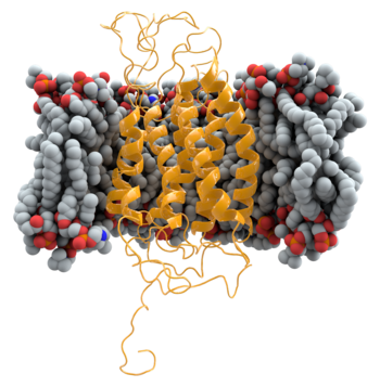
CCR5 is a human membrane protein that acts as a secondary receptor for HIV, enabling the viral and cell membranes to fuse. People with two copies of a mutated Δ32 form of CCR5 are naturally resistant to infection by most strains of HIV, and the normal form is the target of entry inhibitors such as maraviroc. Credit: Thomas Splettstoesser (18 July 2012) |
| Portal:Viruses/Selected picture/26
Megavirus chilensis is a very large DNA virus discovered in 2010. Until the discovery of Pandoravirus in 2013, it was the largest known virus, with its 440 nm diameter capsid being as large as some small bacteria. The capsid is enclosed in bacterial-like capsular material 75–100 nm thick. Credit: Chantal Abergel (10 October 2011) |
| Portal:Viruses/Selected picture/27
Haemagglutinin, a glycoprotein trimer on the influenza virus envelope, binds to the sialic acid-containing receptor on the host cell. After the virus has been engulfed into an endosome, it changes configuration, causing the viral and endosomal membranes to fuse, releasing the viral genome into the cytoplasm. Credit: Jawahar Swaminathan (EBI) (16 November 2008) |
| Portal:Viruses/Selected picture/28
Muhammad ibn Zakariya al-Razi was a Persian physician and chemist who, in the 9th century, was the first to document the distinction between the diseases of measles and smallpox. Credit: Gerard of Cremona (c. 1250–60) |
| Portal:Viruses/Selected picture/29
The castor bean tick, Ixodes ricinus, can transmit the tick-borne encephalitis virus. Ticks are common vectors for viruses, and other tick-borne diseases include Colorado tick fever and Crimean–Congo haemorrhagic fever. Credit: Richard Bartz (24 April 2009) |
| Portal:Viruses/Selected picture/30
Eastern equine encephalitis virus is an Alphavirus that is transmitted between birds and mammals, including humans and horses, by several mosquito species. The virus (coloured in red) is found in the mosquito salivary gland, and is injected into the new host when the insect feeds. Credit: Fred Murphy, Sylvia Whitfield, CDC (1968) |
| Portal:Viruses/Selected picture/31
The striping caused by tulip breaking virus, first described in 1576 by Carolus Clusius, was the second plant virus disease to be documented. The effects were much prized by 17th-century tulip growers. Credit: Unknown (before 1640) |
| Portal:Viruses/Selected picture/32
Ebola virus (coloured green), a filamentous RNA virus, budding from a chronically infected African green monkey kidney cell in culture. Credit: BernbaumJG (28 August 2014) |
| Portal:Viruses/Selected picture/33
False-coloured scanning electron micrograph of severe acute respiratory syndrome coronavirus 2 (SARS-CoV-2; gold) emerging from cells in culture. SARS-CoV-2 is a novel coronavirus first identified in China in January 2020 as the cause of an acute respiratory disease now known as COVID-19. Cases of the disease increased rapidly and spread globally, with the World Health Organization characterising COVID-19 as a pandemic in March 2020. Credit: National Institute of Allergy and Infectious Diseases (19 February 2020) |
| Portal:Viruses/Selected picture/34
False-coloured transmission electron micrograph of severe acute respiratory syndrome coronavirus 2, a novel coronavirus, showing the spikes (blue) forming a crown that give this group of RNA viruses their name. The spike protein interacts with the cellular receptor for the virus, angiotensin-converting enzyme 2. Credit: National Institute of Allergy and Infectious Diseases (13 February 2020) |
| Portal:Viruses/Selected picture/35 |


