Pinworm (parasite)

The pinworm (species Enterobius vermicularis), also known as threadworm (in the United Kingdom, Australia and New Zealand) or seatworm, is a parasitic worm. It is a nematode (roundworm) and a common intestinal parasite or helminth, especially in humans.[7] The medical condition associated with pinworm infestation is known as pinworm infection (enterobiasis)[8] (a type of helminthiasis) or less precisely as oxyuriasis in reference to the family Oxyuridae.[9]
Other than human, Enterobius vermicularis were reported from bonnet macaque.[10] Other species seen in primates include Enterobius buckleyi in Orangutan[11] and Enterobius anthropopitheci in chimpanzee. Enterobius vermicularis is common in human children and transmitted via the faecal-oral route. Humans are the only natural host of Enterobius vermicularis.[12] Enterobius gregorii, another human species is morphologically indistinguishable from Enterobius vermicularis except the spicule size.[13] Throughout this article, the word "pinworm" refers to Enterobius. In British usage, however, pinworm refers to Strongyloides, while Enterobius is called threadworm.[14]
A fossilized nematode egg was detected in 240 million-year-old fossil dung,[15] showing that parasitic pinworms already infested pre-mammalian cynodonts. The earliest known instance of the pinworms associated with humans is evidenced by pinworm eggs found in human coprolites carbon dated to 7837 BC found in western Utah.[16]
Morphology
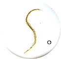
The adult female has a sharply pointed posterior end, is 8 to 13 mm long, and 0.5 mm thick.[17] The adult male is considerably smaller, measuring 2 to 5 mm long and 0.2 mm thick, and has a curved posterior end.[17] The eggs are translucent[17] and have a surface that adheres to objects.[16] The eggs measure 50 to 60 μm by 20 to 30 μm, and have a thick shell flattened on one side.[17] The small size and colourlessness of the eggs make them invisible to the naked eye, except in barely visible clumps of thousands of eggs. Eggs may contain a developing embryo or a fully developed pinworm larva.[17] The larvae grow to 140–150 μm in length.[16]
-

Two female pinworms next to a ruler: The markings are 1 mm apart.
-
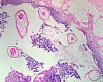
Pinworms are sometimes diagnosed incidentally by pathology. Micrograph of pinworms in the appendix, H&E stain
-
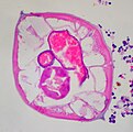
High magnification micrograph of a pinworm in cross section in the appendix, H&E stain
-

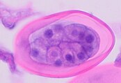
Partially longitudinal cross-section of Enterobius vermicularis, H&E stain
-
Cross-section of early E. vermicularis egg, H&E stain
-

Later E. vermicularis eggs, of the same size as early eggs but having undergone more mitoses. H&E stain
-

Micrograph of male pinworm in cross section, alae (blue arrow), intestine (red arrow) and testis (black arrow), H&E stain
-

Pinworm eggs are easily seen under a microscope.
-
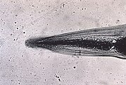
This micrograph reveals the cephalic alae in the head region of E. vermicularis.
-
E. vermicularis

.jpg)
.jpg)


Life cycle

The entire life cycle, from egg to adult, takes place in the human gastrointestinal tract of a single host,[17][16] from about 2–4 weeks[18] or about 4–8 weeks.[19] E. vermicularis molts four times; the first two within the egg before hatching and two before becoming an adult worm.[20]
Although infection often occurs via ingestion of embryonated eggs by inadequate hand washing or nail biting, inhalation followed by swallowing of airborne eggs may occur rarely.[16][19] The eggs hatch in the duodenum (i.e., first part of the small intestine).[21] The emerging pinworm larvae grow rapidly to a size of 140 to 150 μm,[18] and migrate through the small intestine towards the colon.[16] During this migration, they moult twice and become adults.[16][19] Females survive for 5 to 13 weeks, and males about 7 weeks.[16] The male and female pinworms mate in the ileum (i.e., last part of the small intestine),[16] whereafter the male pinworms usually die,[21] and are passed out with stool.[22] The gravid female pinworms settle in the ileum, caecum (i.e., beginning of the large intestine), appendix and ascending colon,[16] where they attach themselves to the mucosa[19] and ingest colonic contents.[23]
Almost the entire body of a gravid female becomes filled with eggs.[21] The estimations of the number of eggs in a gravid female pinworm range from about 11,000[16] to 16,000.[19] The egg-laying process begins about five weeks after initial ingestion of pinworm eggs by the human host.[16] The gravid female pinworms migrate through the colon towards the rectum at a rate of 12 to 14 cm per hour.[16] They emerge from the anus, and while moving on the skin near the anus, the female pinworms deposit eggs either through (1) contracting and expelling the eggs, (2) dying and then disintegrating, or (3) bodily rupture due to the host scratching the worm.[21] After depositing the eggs, the female becomes opaque and dies.[22] The female emerges from the anus to obtain the oxygen necessary for the maturation of the eggs.[22]
Classification
The pinworm (genus Enterobius) is a type of roundworm (nematode), and three species of pinworm have been identified with certainty.[24] Humans are hosts only to Enterobius vermicularis (formerly Oxyurias vermicularis).[25] Chimpanzees are host to Enterobius anthropopitheci, which is morphologically distinguishable from the human pinworm.[5] Hugot (1983) claims another species affects humans, Enterobius gregorii, which is supposedly a sister species of E. vermicularis, and has a slightly smaller spicule (i.e., sexual organ).[26] Its existence is controversial, however; Totkova et al. (2003) consider the evidence to be insufficient,[6] and Hasegawa et al. (2006) contend that E. gregorii is a younger stage of E. vermicularis.[4][5] Regardless of its status as a distinct species, E. gregorii is considered clinically identical to E. vermicularis.[25]
Human infection

E. vermicularis causes the medical condition enterobiasis, whose primary symptom is itching in the anal area.[27] Extraintestinal disease is rare and most commonly involves the female reproductive tract,[28] but spleen abscess has also been reported.[29]
Prevalence
The pinworm has a worldwide distribution,[23] and is the most common helminth (i.e., parasitic worm) infection in the United States, western Europe, and Oceania.[19] In the United States, a study by the Center of Disease Control reported an overall incidence rate of 11.4% among children.[19] Pinworms are particularly common in children, with prevalence rates in this age group having been reported as high as 61% in India, 50% in England, 39% in Thailand, 37% in Sweden, and 29% in Denmark.[19] Finger sucking has been shown to increase both incidence and relapse rates,[19] and nail biting has been similarly associated.[30] Because it spreads from host to host through contamination, pinworms are common among people living in close contact, and tends to occur in all people within a household.[23] The prevalence of pinworms is not associated with gender,[23] nor with any particular social class, race, or culture.[19] Pinworms are an exception to the tenet that intestinal parasites are uncommon in affluent communities.[19]
See also
Notes
- ↑ 1.0 1.1 Hasegawa et al. 2005.
- ↑ 2.0 2.1 2.2 2.3 2.4 2.5 "Enterobius". NCBI taxonomy. Bethesda, MD: National Center for Biotechnology Information. Archived from the original on 1 March 2019. Retrieved 28 February 2019.
- ↑ 3.00 3.01 3.02 3.03 3.04 3.05 3.06 3.07 3.08 3.09 3.10 3.11 3.12 3.13 3.14 3.15 3.16 3.17 3.18 3.19 3.20 3.21 3.22 "GBIF Backbone Taxonomy". GBIF Secretariat. 2022. doi:10.15468/39omei. Archived from the original on 1 February 2022. Retrieved 16 September 2023.
{{cite journal}}: Cite journal requires|journal=(help) - ↑ 4.0 4.1 Hasegawa et al. 1998
- ↑ 5.0 5.1 5.2 Hasegawa et al. 2006
- ↑ 6.0 6.1 Totkova et al. 2003
- ↑ Encyclopædia Britannica.
- ↑ Merriam-Webster: Enterobiasis
- ↑ Merriam-Webster: Oxyuriasis
- ↑ C.P., Arjun (October 2015). "A Study of Gastrointestinal Parasites in Bonnet Macaques (Macaca radiata) of Pookode, Wayanad, Kerala". Zoos' Print Journal. Zoo Outreach Organization. Archived from the original on 1 April 2016. Retrieved 20 October 2015.
- ↑ Foitová, Ivona; Civáňová, Kristína; Baruš, Vlastimil; Nurcahyo, Wisnu (1 July 2014). "Phylogenetic relationships between pinworms (Nematoda: Enterobiinae) parasitising the critically endangered orang-utan, according to the characterisation of molecular genomic and mitochondrial markers". Parasitology Research. 113 (7): 2455–2466. doi:10.1007/s00436-014-3892-y. ISSN 1432-1955. PMID 24880237. S2CID 15076891.
- ↑ Panidis, Stavros; Paramythiotis, Daniel; Panagiotou, Dimitris; Batsis, Georgios; Salonikidis, Spyridon; Kaloutsi, Vassiliki; Michalopoulos, Antonios (1 January 2011). "Acute appendicitis secondary to Enterobius vermicularis infection in a middle-aged man: a case report". Journal of Medical Case Reports. 5: 559. doi:10.1186/1752-1947-5-559. ISSN 1752-1947. PMC 3245485. PMID 22128765.
- ↑ CP, Arjun (October 2015). "A Study of Gastrointestinal Parasites in Bonnet Macaque ( Macaca radiata) of Pookode, Wayanad, Kerala" (PDF). Zoos' Print Journal. 10. Archived (PDF) from the original on 21 August 2016. Retrieved 16 September 2023.
- ↑ Vanderkooi 2000, p. B-152 & B-225
- ↑ Mitrica, Dragos (2 December 2014). "Scientists find 240 million-year-old parasite that infected mammals' ancestor". ZME Science. Archived from the original on 14 February 2023. Retrieved 14 February 2023.
- ↑ 16.00 16.01 16.02 16.03 16.04 16.05 16.06 16.07 16.08 16.09 16.10 16.11 16.12 Cook 1994, p. 1159
- ↑ 17.0 17.1 17.2 17.3 17.4 17.5 Gutiérrez 2005, p. 354.
- ↑ 18.0 18.1 Cook et al. 2009, p. 1516
- ↑ 19.00 19.01 19.02 19.03 19.04 19.05 19.06 19.07 19.08 19.09 19.10 Burkhart & burkhart 2005, p. 837
- ↑ Rett, Doug. "Enterobius vermicularis". Animal Diversity Web. Archived from the original on 9 June 2023. Retrieved 4 January 2021.
- ↑ 21.0 21.1 21.2 21.3 Garcia 1999, p. 246
- ↑ 22.0 22.1 22.2 Caldwell 1982, p. 307.
- ↑ 23.0 23.1 23.2 23.3 Gutiérrez 2005, p. 355.
- ↑ NCBI taxonomy database 2009
- ↑ 25.0 25.1 dpdx 2009
- ↑ Hugot 1983
- ↑ "Enterobiasis leads to itching". Archived from the original on 19 September 2023. Retrieved 20 August 2011.
- ↑ Graves, B.; Leder, K.; Sinickas, V.; Sheorey, H. (February 2018). "Extraintestinal Enterobius vermicularis". Pathology. 50: S113–S114. doi:10.1016/j.pathol.2017.12.322. Archived from the original on 19 September 2023. Retrieved 16 September 2023.
- ↑ Agostinis, Paolo; Cappello, Dario; Riccardi, Niccolò; Michelutti, Teresa; Orsaria, Maria; Zerbato, Verena; Di Bella, Stefano (11 September 2023). Amoroso, Anthony; Nori, Priya; Riedel, David J (eds.). "A 25-Year-Old Woman With Long-Lasting Abdominal Pain and Spleen Abscess". Clinical Infectious Diseases. 77 (5): 795–798. doi:10.1093/cid/ciad047. ISSN 1058-4838. Archived from the original on 19 September 2023. Retrieved 16 September 2023.
- ↑ Cook 1994, p. 1160
References
- Hasegawa H, Ikeda Y, Fujisaki A, et al. (December 2005). "Morphology of chimpanzee pinworms, Enterobius (Enterobius) anthropopitheci (Gedoelst, 1916) (Nematoda: Oxyuridae), collected from chimpanzees, Pan troglodytes, on Rubondo Island, Tanzania". The Journal of Parasitology. 91 (6): 1314–7. doi:10.1645/GE-569R.1. PMID 16539010. S2CID 32110983.
- "Pinworm". Encyclopædia Britannica. Archived from the original on 15 May 2015. Retrieved 8 April 2009.
- "Enterobiasis". Merriam-Webster's Medical Dictionary. Merriam-Webster. Archived from the original on 3 March 2016. Retrieved 8 April 2009.
- "Oxyuriasis". Merriam-Webster's Medical Dictionary. Merriam-Webster. Archived from the original on 24 February 2021. Retrieved 8 April 2009.
- Totkova A, Klobusicky M, Holkova R, Valent M (2003). "Enterobius gregorii—reality or fiction?" (PDF). Bratislavské Lekárske Listy. 104 (3): 130–3. PMID 12940699. Archived from the original (PDF) on 10 September 2011.
- "Enterobius". NCBI taxonomy database. National Center for Biotechnology Information, U.S. National Library of Medicine. 2009. Archived from the original on 14 May 2018. Retrieved 8 April 2009.
- "Enterobiasis". DPDx. Division of Parasitic Diseases, Centers for Disease Control and Prevention. Archived from the original on 27 November 2013. Retrieved 8 April 2009.
- Nakano T, Okamoto M, Ikeda Y, Hasegawa H (December 2006). "Mitochondrial cytochrome c oxidase subunit 1 gene and nuclear rDNA regions of Enterobius vermicularis parasitic in captive chimpanzees with special reference to its relationship with pinworms in humans". Parasitology Research. 100 (1): 51–7. doi:10.1007/s00436-006-0238-4. PMID 16788831. S2CID 32762371.
- Hugot JP (1983). "[Enterobius gregorii (Oxyuridae, Nematoda), a new human parasite]". Annales de Parasitologie Humaine et Comparée (in français). 58 (4): 403–4. doi:10.1051/parasite/1983584403. PMID 6416131.
- Hasegawa H, Takao Y, Nakao M, Fukuma T, Tsuruta O, Ide K (February 1998). "Is Enterobius gregorii Hugot, 1983 (Nematoda: Oxyuridae) a distinct species?". The Journal of Parasitology. 84 (1): 131–4. doi:10.2307/3284542. JSTOR 3284542. PMID 9488350.
- Gutiérrez, Yezid (2000). Diagnostic pathology of parasitic infections with clinical correlations (Second ed.). Oxford University Press. pp. 354–366. ISBN 978-0-19-512143-8. Archived from the original on 19 September 2023. Retrieved 16 September 2023.
- Cook, Gordon C; Zumla, Alimuddin I (2009). Manson's tropical diseases (22nd ed.). Saunders Elsevier. pp. 1515–1519. ISBN 978-1-4160-4470-3. Archived from the original on 19 September 2023. Retrieved 16 September 2023.
- "B80: Enterobiasis". International Statistical Classification of Diseases and Related Health Problems (ICD) 10th Revision. World Health Organization. 2007. Archived from the original on 6 September 2009. Retrieved 5 December 2009.
- Cook GC (September 1994). "Enterobius vermicularis infection". Gut. 35 (9): 1159–62. doi:10.1136/gut.35.9.1159. PMC 1375686. PMID 7959218.
- Garcia, Lynne Shore (2009). Practical guide to diagnostic parasitology. American Society for Microbiology. pp. 246–247. ISBN 978-1-55581-154-9. Archived from the original on 19 September 2023. Retrieved 16 September 2023.
- Burkhart CN, Burkhart CG (October 2005). "Assessment of frequency, transmission, and genitourinary complications of enterobiasis (pinworms)". International Journal of Dermatology. 44 (10): 837–40. doi:10.1111/j.1365-4632.2004.02332.x. PMID 16207185. S2CID 3193432.
- Caldwell JP (February 1982). "Pinworms (Enterobius Vermicularis)". Canadian Family Physician. 28: 306–9. PMC 2306321. PMID 21286054.
- Vanderkooi M (2000). Village Medical Manual (5th ed.).
External links
- CS1 errors: missing periodical
- Articles with hatnote templates targeting a nonexistent page
- Articles with redirect hatnotes needing review
- Use dmy dates from May 2021
- Articles with invalid date parameter in template
- Articles with 'species' microformats
- CS1 français-language sources (fr)
- Oxyurida
- Parasitic nematodes of humans
- Parasitic nematodes of mammals
- Parasites of equines
- Parasites of primates
- Colorectal surgery