Pemphigus vulgaris
| Pemphigus vulgaris | |
|---|---|
.jpg) | |
| Specialty | Dermatology |
Pemphigus vulgaris is a rare chronic blistering skin disease and the most common form of pemphigus. Pemphigus was derived from the Greek word pemphix, meaning blister.[1] It is classified as a type II hypersensitivity reaction in which antibodies are formed against desmosomes, components of the skin that function to keep certain layers of skin bound to each other. As desmosomes are attacked, the layers of skin separate and the clinical picture resembles a blister. These blisters are due to acantholysis, or breaking apart of intercellular connections through an autoantibody-mediated response.[2] Over time the condition inevitably progresses without treatment: lesions increase in size and distribution throughout the body, behaving physiologically like a severe burn.
Before the advent of modern treatments, mortality for the disease was close to 90%. Today, the mortality rate with treatment is between 5-15% due to the introduction of corticosteroids as primary treatment.[3] Nevertheless, in 1998, pemphigus vulgaris was the fourth most common cause of death due to a skin disorder.
The disease mainly affects middle-aged and older adults between 50–60 years old. There has historically been a higher incidence in women.[4]
Signs and symptoms
Pemphigus vulgaris most commonly presents with oral blisters (buccal and palatine mucosa, especially), but also includes cutaneous blisters. Other mucosal surfaces, the conjunctiva, nose, esophagus, penis, vulva, vagina, cervix, and anus, may also be affected. Flaccid blisters over the skin are frequently seen with sparing of the skin covering the palms and soles.[5]
Blisters commonly erode and leave ulcerated lesions and erosions. A positive Nikolsky sign (induction of blistering in normal skin or at the edge of a blister) is indicative of the disease.[5]
Severe pain with chewing can lead to weight loss and malnutrition.[5]
-
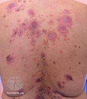
Pemphigus vulgaris
-
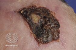
Pemphigus vulgaris
-

Pemphigus vulgaris
-
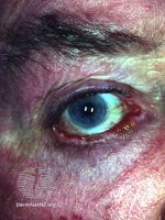
Ocular pemphigus vulgaris
.jpg)
.jpg)

.jpg)
Pathophysiology
Pemphigus is an autoimmune disease caused by antibodies directed against both desmoglein 1 and desmoglein 3 present in desmosomes. Loss of desmosomes results in loss of cohesion between keratinocytes in the epidermis, and a disruption of the barrier function served by intact skin. The process is classified as a type II hypersensitivity reaction (in which antibodies bind to antigens on the body's own tissues). On histology, the basal keratinocytes are usually still attached to the basement membrane leading to a characteristic appearance called "tombstoning". Transudative fluid accumulates in between the keratinocytes and the basal layer (suprabasal split), forming a blister and resulting in what is known as a positive Nikolsky's sign. This is a contrasting feature from bullous pemphigoid, which is thought to be due to anti-hemidesmosome antibodies, and where the detachment occurs between the epidermis and dermis (subepidermal bullae). Clinically, pemphigus vulgaris is characterized by extensive flaccid blisters and mucocutaneous erosions. The severity of the disease, as well as the mucosal lesions, is believed to be directly proportional to the levels of desmoglein 3. Milder forms of pemphigus (like foliacious and erythematoses) are more anti-desmoglein 1 heavy.
The disease arises most often in middle-aged or older people, usually starting with a blister that ruptures easily. It can also start with blisters in the mouth. The lesions can become quite extensive.
Diagnosis

Because it is a rare disease, diagnosis is often complicated and takes a long time. Early in the disease patients may have erosions in the mouth or blisters on the skin. These blisters can be itchy or painful. Theoretically, the blisters should demonstrate a positive Nikolsky's sign, in which the skin sloughs off from slight rubbing, but this is not always reliable. The gold standard for diagnosis is a punch biopsy from the area around the lesion that is examined by direct immunofluorescent staining, in which cells are acantholytic, that is, lacking the normal intercellular connections that hold them together. These can also be seen on a Tzanck smear. These cells are basically rounded, nucleated keratinocytes formed due to antibody mediated damage to cell adhesion protein desmoglein.
Pemphigus vulgaris is easily confused with impetigo and candidiasis. IgG4 is considered pathogenic. The diagnosis can be confirmed by testing for the infections that cause these other conditions, and by a lack of response to antibiotic treatment.[6]
Treatment
Corticosteroids and other immunosuppressive medications have historically been employed to reduce pemphigus symptoms, yet steroids are associated with serious and long-lasting side effects and their use should be limited as much as possible. Intravenous immunoglobulin, mycophenolate mofetil, methotrexate, azathioprine, and cyclophosphamide have also been used with varying degrees of success.
An established alternative to steroids are monoclonal antibodies such as rituximab, which are increasingly being used as first-line treatment. In summer 2018, the FDA granted full approval to rituximab for this application, following successful fast track evaluation.[7] In numerous case series, many patients achieve remission after one cycle of rituximab. Treatment is more successful if initiated early on in the course of disease, perhaps even at diagnosis. Rituximab treatment combined with monthly IV immunoglobulin infusions has resulted in long-term remission with no recurrence of disease in 10 years after treatment was halted.[8] This was a small trial study of 11 patients with 10 patients followed to completion.
Epidemiology
Pemphigus vulgaris is a relatively rare disease that only affects about 1 to 5 people in 1 million in the United Kingdom, with an incidence of 1-10 cases per 1 million people across the world. There is an estimated prevalence of 30,000-40,000 cases in the United States.[9] Cases of P. vulgaris usually don't develop until after the age of 50 or so. The disease is not contagious which means it cannot be spread from person to person.[10] There is currently no way of knowing who will be affected with P. vulgaris in their life as it is usually not a genetic disorder and is usually triggered later in life by environmental factors. Men and women are both equally affected and the disease has been found to affect people of many different cultures and racial backgrounds, especially Ashkenazi Jews, people of Mediterranean, North Indian and Persian descent.[11] There has been no found difference in the rate of disease when looking at socioeconomic factors as well.[9]
If left untreated, 8 of 10 people with the disease die within a year with a cause of death being infection or loss of fluids, which is very common for raw, open sores that are characteristic of P. vulgaris. With treatment, only about 1 in 10 people with the disease die, either from the condition, or side effects of the medicine.[10]
An effect of the disease being so rare is that there is not enough evidence to prove that the treatments currently being used are actually as effective as they could be. Doctors are trying to find effective steroid-sparing agents to use in the treatment, to decrease the side effects of long term steroid treatment. The small amount of case numbers make it hard to test statistical significance between the affected and the control groups when testing if these types of systematic treatments are effective.[12]
Research
Research into using genetically modified T-cells to treat pemphigus vulgaris in mice was reported in 2016.[13][14] Rituximab indiscriminately attacks all B cells, which reduces the body's ability to control infections. In the experimental treatment, human T cells are genetically engineered to recognize only those B cells that produce antibodies to desmoglein. In PV, autoreactive B cells produce antibodies against Dsg3, disrupting its adhesive function and causing skin blistering. By expressing Dsg3 on their surfaces, the CAAR-T cells lure those B cells in and kill them. The Dsg 3 CAAR-T cells eliminated Dsg3-specific B cells in lab dishes and in mice, the researchers reported at the time. Penn's Aimee Payne, M.D., Ph.D., and Michael Milone, M.D., Ph.D., a co-inventor of Novartis’ CAR-T cancer therapy Kymriah, pioneered the CAAR-T idea and have founded biotech Cabaletta Bio, hoping to bring their therapies through clinical studies to the market. The MuSK-CAAR-T is the second candidate in its pipeline; the company has already received FDA clearance to test its lead project, DSG3-CAAR-T, in patients with mucosal pemphigus vulgaris (PV), and a phase 1 trial is expected to start in 2020.
See also
- List of conditions caused by problems with junctional proteins
- List of cutaneous conditions
- List of immunofluorescence findings for autoimmune bullous conditions
References
- ↑ Cholera M, Chainani-Wu N (June 2016). "Management of Pemphigus Vulgaris". Advances in Therapy. 33 (6): 910–58. doi:10.1007/s12325-016-0343-4. PMC 4920839. PMID 27287854.
- ↑ Beigi PK (2018). "History of Pemphigus Vulgaris". A Clinician's Guide to Pemphigus Vulgaris. Springer, Cham. p. 13. doi:10.1007/978-3-319-67759-0_3. ISBN 978-3-319-67758-3.
- ↑ King DF, Holubar K (October 1983). "History of pemphigus". Clinics in Dermatology. 1 (2): 6–12. doi:10.1016/0738-081X(83)90019-6. PMID 6400552.
- ↑ Gupta VK, Kelbel TE, Nguyen D, Melonakos KC, Murrell DF, Xie Y, Mullard A, Reed PL, Seiffert-Sinha K, Sinha AA (July 2011). "A globally available internet-based patient survey of pemphigus vulgaris: epidemiology and disease characteristics". Dermatologic Clinics. 29 (3): 393–404, vii–iii. doi:10.1016/j.det.2011.03.016. PMID 21605804.
- ↑ 5.0 5.1 5.2 "Pathogenesis, clinical manifestations, and diagnosis of pemphigus". www.uptodate.com. Archived from the original on 2017-04-04. Retrieved 2017-04-03.
- ↑ Kaplan DL (2009). "Dermclinic". Consultant. 49 (3). Archived from the original on 2012-02-13. Retrieved 2020-12-22.
- ↑ "FDA Approves Genentech's Rituxan (Rituximab) for Pemphigus Vulgaris". Genentech. Archived from the original on 2019-12-24. Retrieved 24 December 2019.
- ↑ Ahmed AR, Kaveri S, Spigelman Z (December 2015). "Long-Term Remissions in Recalcitrant Pemphigus Vulgaris". The New England Journal of Medicine. 373 (27): 2693–4. doi:10.1056/NEJMc1508234. PMID 26716930.
- ↑ 9.0 9.1 "Pemphigus". www.pemphigus.org. Archived from the original on 2019-12-07. Retrieved 2019-12-03.
- ↑ 10.0 10.1 "Pemphigus Vulgaris | Causes, Symptoms and Treatment". patient.info. Archived from the original on 2019-06-22. Retrieved 2019-11-18.
- ↑ "Pemphigus and Pemphigoid". NORD (National Organization for Rare Disorders). Archived from the original on 2017-03-12. Retrieved 2019-11-18.
- ↑ Daniel, Benjamin S.; Murrell, Dedee F. (2014-05-06). "Management of pemphigus". F1000Prime Reports. 6: 32. doi:10.12703/P6-32. ISSN 2051-7599. PMC 4017903. PMID 24860654.
- ↑ Gallagher J (2016-07-01). "'Civil war' in immune system can fight disease". BBC News, Health. Archived from the original on 2016-07-01. Retrieved 2016-07-01.
- ↑ Ellebrecht CT, Bhoj VG, Nace A, Choi EJ, Mao X, Cho MJ, Di Zenzo G, Lanzavecchia A, Seykora JT, Cotsarelis G, Milone MC, Payne AS (July 2016). "Reengineering chimeric antigen receptor T cells for targeted therapy of autoimmune disease". Science. 353 (6295): 179–84. Bibcode:2016Sci...353..179E. doi:10.1126/science.aaf6756. PMC 5343513. PMID 27365313.
External links
| Classification | |
|---|---|
| External resources |
- Pemphigus vulgaris - DermNet New Zealand Archived 2020-11-29 at the Wayback Machine