Mycosis fungoides
| Mycosis fungoides | |
|---|---|
| Other names: Alibert-Bazin syndrome, granuloma fungoides[1] | |
 | |
| Skin lesions on the knee of a 52-year-old male patient with Mycosis fungoides | |
| Specialty | Oncology, dermatology |
| Causes | Unknown[2] |
| Risk factors | Organ transplantation, long-term exposure to allergens, runs in some families[2] |
| Frequency | 5-6 per million people, males>females, elderly>children, Black>white[2] |
Mycosis fungoides is the most common form of cutaneous T-cell lymphoma, a type of skin tumor which has several variants.[2] It generally affects the skin, but may progress internally over time. Symptoms include rash, tumors, skin lesions, and itchy skin.
While the cause remains unclear, most cases are not hereditary. Most cases are in people over 20 years of age, and it is more common in men than women. Treatment options include sunlight exposure, ultraviolet light, topical corticosteroids, chemotherapy, and radiotherapy.
Signs and symptoms
The symptoms of mycosis fungoides are categorized into three clinical stages: the patch stage, the plaque stage, and the tumour stage.[3] The patch stage is defined by flat, reddish patches of varying sizes that may have a wrinkled appearance. They can also look yellowish in people with darker skin.[3] The plaque stage follows the patch stage of mycosis fungoides.[4] It is characterized by the presence of raised lesions that appear reddish-brown; in darker skin tones, plaques may have a greyish or silver appearance.[5] Both patch and plaque stages are considered early-stage mycosis fungoides.[4] The tumour stage typically shows large irregular lumps. Tumours can develop form plaques or normal skin in any region of the body, including the face and head regions.[6]
Itching (pruritus) is common, perhaps in 20 percent of patients. The symptoms displayed are progressive, with early stages consisting of lesions presented as scaly patches. Lesions often initially develop on the trunk of the body in places that are rarely exposed to the sun, such as the buttocks.[3] The advanced stage of mycosis fungoides is characterized by generalized erythroderma (red rash covering most of the body) with severe pruritus (itching) and scaling.[5] The key difference between Sézary syndrome and the other stages of mycosis fungoides is the large number of cancer cells found in the blood (leukemic disease).[7] When mycosis fungoides develops to meet the criteria for Sézary syndrome, it is referred to as leukemic mycosis fungoides, Sézary syndrome preceded by mycosis fungoides, or secondary mycosis fungoides.[7]
-
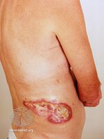
Ulcerated tumour stage mycosis fungoides
-
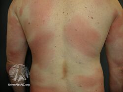
Patch stage mycosis fungoides
-
Patch stage mycosis fungoides
-

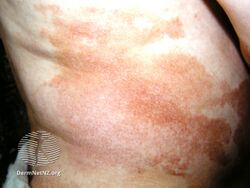
Plaque of mycosis fungoides
.jpg)
.jpg)
.jpg)

Cause
The cause of mycosis fungoides is unknown. A current hypothesized cause is genetic and cellular signalling abnormalities. Examples of these abnormalities include clonal abnormalities and altered expression of adhesion molecules and/or cytokins.[8]
Additionally, the disease is an unusual expression of CD4 T cells, a part of the immune system. These T cells are skin-associated, meaning they are biochemically and biologically most related to the skin, in a dynamic manner. Mycosis fungoides is the most common type of cutaneous T-cell lymphoma (CTCL), but there are many other types of CTCL that have nothing to do with mycosis fungoides and these disorders are treated differently.[9]
Diagnosis
Diagnosis is sometimes difficult because the early phases of the disease often resemble inflammatory dermatoses (such as eczema, psoriasis, lichenoid dermatoses including lichen planus, vitiligo, and chronic cutaneous lupus erythematosus), as well as other cutaneous lymphomas.[10] Several biopsies are recommended, to be more certain of the diagnosis.
The criteria for the disease are established on the skin biopsy:[11]
- the superficial papillary dermis is infiltrated by a bandlike lymphocyte infiltrate
- epidermotropism
- presence of atypical T-cells with cerebriform nuclei in the dermal and epidermal infiltrates.

Pautrier's microabcesses are aggregates of four or more atypical lymphocytes arranged in the epidermis.[12] Pautrier microabcesses are characteristic of mycosis fungoides but are generally absent.
To stage the disease, various tests may be ordered, to assess nodes, blood and internal organs, but most patients present with disease apparently confined to the skin, as patches (flat spots) and plaques (slightly raised or 'wrinkled' spots).
Peripheral smear will often show buttock cells.[13]
Staging
Traditionally, mycosis fungoides has been divided into three stages: premycotic, mycotic and tumorous. The premycotic stage clinically presents as an erythematous (red), itchy, scaly lesion. Microscopic appearance is non-diagnostic and represented by chronic nonspecific dermatosis associated with psoriasiform changes in epidermis.[medical citation needed]
In the mycotic stage, infiltrative plaques appear and biopsy shows a polymorphous inflammatory infiltrate in the dermis that contains small numbers of frankly atypical lymphoid cells. These cells may line up individually along the epidermal basal layer. The latter finding if unaccompanied by spongiosis is highly suggestive of mycosis fungoides. In the tumorous stage a dense infiltrate of medium-sized lymphocytes with cerebriform nuclei expands the dermis.
Accurate staging of Mycosis Fungoides is essential to determine appropriate treatment and prognosis.[14] Staging is based on the tumor, node, metastasis, blood (TNMB) classification proposed by the Mycosis Fungoides Cooperative Group and revised by the International Society for Cutaneous Lymphomas/European Organization of Research and Treatment of Cancer.[14] This staging system examines the extent of skin involvement (T), presence of lymph node (N), visceral disease (M), and presence of Sezary cells in the peripheral blood (B).[14]
Most patients with Mycosis Fungoides have early-stage disease (Stage IA-IIA) at the time of their initial diagnosis.[14] These patients with early stage disease that is primarily confined to the skin have a favorable prognosis.[14] Patients with advanced stage (Stage IIB-IVB) are often refractory to treatment and have an unfavorable prognosis.[14] Treatment of patients with advanced stage disease is designed to reduce tumor burden, delay disease progression, and preserve quality of life.[14]
Treatment
Mycosis fungoides can be treated in a variety of ways.[15] Common treatments include simple sunlight, ultraviolet light (mainly NB-UVB 312 nm), topical steroids, topical and systemic chemotherapies, local superficial radiotherapy, the histone deacetylase inhibitor vorinostat, total skin electron radiation, photopheresis and systemic therapies (e.g. interferons, retinoids, rexinoids) or biological therapies.[15] Treatments are often used in combination.[16]
There is limited evidence for the efficacy of topical and systemic therapies for mycosis fungoides.[16] Due to the possible adverse effects of these treatments in early disease it is recommended to begin therapy with topical and skin-directed treatments before progressing to more systemic therapies.[16] Larger and more extensive research is needed to identify effective treatment strategies for this disease.[16]
In 2010, the U.S. Food and Drug Administration granted orphan drug designation for naloxone lotion, a topical opioid receptor competitive antagonist used as a treatment for pruritus in cutaneous T-cell lymphoma.[17][18]
The US FDA approved the drug mogamulizumab (trade name Poteligeo) in August 2018 for treatment of relapsed or refractory mycosis fungoides and Sézary disease.[19]
Prognosis
A 1999 US-based study of patient records observed a 5-year relative survival rate of 77%, and a 10-year relative survival rate of 69%. After 11 years, the observed relative survival rate remained around 66%. Poorer survival is correlated with advanced age and black race. Superior survival was observed for married women compared with other gender and marital-status groups.[20]
Epidemiology
It is rare for the disease to appear before age 20, and it appears to be noticeably more common in males than females, especially over the age of 50, where the incidence of the disease (the risk per person in the population) does increase. The average age of onset is between 45 and 55 years of age for patients with patch and plaque disease only, but is over 60 for patients who present with tumours, erythroderma (red skin) or a leukemic form (the Sézary syndrome). The incidence of mycosis fungoides was seen to be increasing till the year 2000 in the United States,[21] thought to be due to improvements in diagnostics. However, the reported incidence of the disease has since then remained constant,[22] suggesting another unknown reason for the jump seen before 2000.
History
Mycosis fungoides was first described in 1806 by French dermatologist Jean-Louis-Marc Alibert.[23][24] The name mycosis fungoides is very misleading—it loosely means "mushroom-like fungal disease". The disease, however, is not a fungal infection but rather a type of non-Hodgkin's lymphoma. It was so named because Alibert described the skin tumors of a severe case as having a mushroom-like appearance.[25]
Culture and society
Notable cases
In 1995 actor Mr. T was diagnosed with a cutaneous T-cell lymphoma, or mycosis fungoides.[26] Once in remission, he joked about the coincidence: "Can you imagine that? Cancer with my name on it — personalized cancer![27]"
Popular British television actor Paul Eddington died of mycosis fungoides after living with the condition for four decades.[28]
See also
- Cutaneous T-cell lymphoma
- Pagetoid reticulosis
- Premycotic phase
- Sézary's disease
- Secondary cutaneous CD30+ large cell lymphoma
- Angiocentric lymphoma
- List of cutaneous conditions
References
- ↑ synd/98 at Who Named It?
- ↑ 2.0 2.1 2.2 2.3 DE, Elder; D, Massi; RA, Scolyer; R, Willemze (2018). "Tumors of haemopoietic and lymphoid origin: Mycosis fungoides". WHO Classification of Skin Tumours. Vol. 11 (4th ed.). Lyon (France): World Health Organization. pp. 226–230. ISBN 978-92-832-2440-2. Archived from the original on 2022-07-11. Retrieved 2022-08-08.
- ↑ 3.0 3.1 3.2 Harvey, Nathan T.; Spagnolo, Dominic V.; Wood, Benjamin A. (2015-12-01). "'Could it be mycosis fungoides?': an approach to diagnosing patch stage mycosis fungoides". Journal of Hematopathology. 8 (4): 209–223. doi:10.1007/s12308-015-0247-2. ISSN 1865-5785.
- ↑ 4.0 4.1 Hristov, Alexandra C.; Tejasvi, Trilokraj; Wilcox, Ryan A. (September 2019). "Mycosis fungoides and Sézary syndrome: 2019 update on diagnosis, risk-stratification, and management". American Journal of Hematology. 94 (9): 1027–1041. doi:10.1002/ajh.25577. ISSN 1096-8652. PMID 31313347. Archived from the original on 2021-08-28. Retrieved 2021-04-24.
- ↑ 5.0 5.1 Cerroni, Lorenzo (March 2018). "Mycosis fungoides-clinical and histopathologic features, differential diagnosis, and treatment". Seminars in Cutaneous Medicine and Surgery. 37 (1): 2–10. doi:10.12788/j.sder.2018.002. ISSN 1085-5629. PMID 29719014. Archived from the original on 2021-08-28. Retrieved 2021-04-24.
- ↑ Valipour, Arash; Jäger, Manuel; Wu, Peggy; Schmitt, Jochen; Bunch, Charles; Weberschock, Tobias (7 July 2020). "Interventions for mycosis fungoides". The Cochrane Database of Systematic Reviews. 7: CD008946. doi:10.1002/14651858.CD008946.pub3. ISSN 1469-493X. PMC 7389258. PMID 32632956. Archived from the original on 18 May 2021. Retrieved 24 April 2021.
{{cite journal}}: CS1 maint: PMC embargo expired (link) - ↑ 7.0 7.1 Larocca, Cecilia; Kupper, Thomas (February 2019). "Mycosis Fungoides and Sézary Syndrome: An Update". Hematology/Oncology Clinics of North America. 33 (1): 103–120. doi:10.1016/j.hoc.2018.09.001. ISSN 1558-1977. PMC 7147244. PMID 30497668. Archived from the original on 2021-08-28. Retrieved 2021-04-24.
- ↑ Khan Mohammad Beigi, Pooya. (2017). Clinician's Guide to Mycosis Fungoides. 10.1007/978-3-319-47907-1_9.
- ↑ Rubio-Gonzalez, B; Zain, J; Rosen, ST; Querfeld, C (January 2017). "Clinical manifestations and pathogenesis of cutaneous lymphomas: current status and future directions". British Journal of Haematology. 176 (1): 16–36. doi:10.1111/bjh.14402. PMID 27782301.
- ↑ Cerroni, L (March 2018). "Mycosis fungoides-clinical and histopathologic features, differential diagnosis, and treatment". Seminars in Cutaneous Medicine and Surgery. 37 (1): 2–10. doi:10.12788/j.sder.2018.002. PMID 29719014.
- ↑ Canada, editors, John P. Greer, MD, Professor, Departments of Medicine and Pediatrics, Divisions of Hermatology/Oncology, Vanderbilt University Medical Center, Nashville, Tennessee; Daniel A. Arber, MD, Professor and Vice Chair, Department of Pathology, Stanford University, Director of Anatomic and Clinical Pathology Services, Stanford University Medical Center, Stanford, California; Bertil Glader, MD, Professor, Departments of Pediatrics and Pathology, Stanford University Medical Center, Stanford, California, Lucile Packard Children's Hospital, Palo Alto, California; Alan F. List, MD, Senior Member, Department of Malignant Hematology, President and CEO, Moffit Cancer Center, Tampa Florida; Robert T. Means Jr., MD, PhD, Professor of Internal Medicine, Executive Dean, University of Kentucky College of Medicine, Lexington, Kentucky; Frixos Paraskevas, MD, Professor of Internal Medicine and Immunology (Retired), University of Manitoba Medical School, Associate Member, Institute of Cell Biology-Cancer Care, Manitoba, Winnipeg, Manitoba, Canada; George M. Rodgers, MD, Professor of Medicine and Pathology, University of Utah School of Medicine, Health Sciences Center, Medical Director, Coagulation Laboratory, ARUB Laboratories, Salt Lake City, Utah; Editor Emeritus, John Foerster, MD, FRCPC, Professor and Physician Emertius, Winnipeg (2014). Wintrobe's clinical hematology (Thirteenth ed.). p. 1957. ISBN 978-1451172683.
- ↑ al.], [edited by] Ronald Hoffman ... [et (2013). Hematology : basic principles and practice (6th ed.). Philadelphia, PA: Saunders/Elsevier. p. 1288. ISBN 978-1437729283.
- ↑ O'Connell, Theodore X. (28 November 2013). USMLE Step 2 Secrets. Elsevier Health Sciences. p. 240. ISBN 9780323225021.
- ↑ 14.0 14.1 14.2 14.3 14.4 14.5 14.6 Jawed, Sarah I.; Myskowski, Patricia L.; Horwitz, Steven; Moskowitz, Alison; Querfeld, Christiane (February 2014). "Primary cutaneous T-cell lymphoma (mycosis fungoides and Sézary syndrome)". Journal of the American Academy of Dermatology. 70 (2): 223.e1–223.e17. doi:10.1016/j.jaad.2013.08.033. PMID 24438970. Archived from the original on 2018-06-30. Retrieved 2021-04-24.
- ↑ 15.0 15.1 Prince HM, Whittaker S, Hoppe RT (November 2009). "How I treat mycosis fungoides and Sézary syndrome". Blood. 114 (20): 4337–53. doi:10.1182/blood-2009-07-202895. PMID 19696197.
- ↑ 16.0 16.1 16.2 16.3 Weberschock, Tobias; Strametz, Reinhard; Lorenz, Maria; Röllig, Christoph; Bunch, Charles; Bauer, Andrea; Schmitt, Jochen (2012-09-12). "Interventions for mycosis fungoides". The Cochrane Database of Systematic Reviews (9): CD008946. doi:10.1002/14651858.CD008946.pub2. ISSN 1469-493X. PMID 22972128.
- ↑ "Elorac, Inc. announces orphan drug designation for novel topical treatment for pruritus in cutaneous T-cell lymphoma (CTCL)". Archived from the original on 2010-12-30. Retrieved 2010-11-30.
- ↑ Wang H, Yosipovitch G (January 2010). "New insights into the pathophysiology and treatment of chronic itch in patients with end-stage renal disease, chronic liver disease, and lymphoma". International Journal of Dermatology. 49 (1): 1–11. doi:10.1111/j.1365-4632.2009.04249.x. PMC 2871329. PMID 20465602.
- ↑ "Press Announcements - FDA approves treatment for two rare types of non-Hodgkin lymphoma". Archived from the original on 2019-04-23. Retrieved 2021-04-24.
- ↑ Weinstock, Martin A.; Reynes, Josefina F. (1999). "The changing survival of patients with mycosis fungoides". Cancer. 85 (1): 208–212. doi:10.1002/(SICI)1097-0142(19990101)85:1<208::AID-CNCR28>3.0.CO;2-2. ISSN 0008-543X.
- ↑ Morales Suárez-Varela, M. M.; Llopis González, A.; Marquina Vila, A.; Bell, J. (2000). "Mycosis fungoides: review of epidemiological observations". Dermatology. 201 (1): 21–28. doi:10.1159/000018423. ISSN 1018-8665. PMID 10971054. S2CID 1544301.
- ↑ Korgavkar, Kaveri; Xiong, Michael; Weinstock, Martin (2013-11-01). "Changing incidence trends of cutaneous T-cell lymphoma". JAMA Dermatology. 149 (11): 1295–1299. doi:10.1001/jamadermatol.2013.5526. ISSN 2168-6084. PMID 24005876.
- ↑ Rapini, Ronald P.; Bolognia, Jean L.; Jorizzo, Joseph L. (2007). Dermatology: 2-Volume Set. St. Louis: Mosby. p. 1867. ISBN 978-1-4160-2999-1.
- ↑ Alibert JLM (1806). Descriptions des maladies de la peau observées a l'Hôpital Saint-Louis, et exposition des meilleures méthodes suivies pour leur traitement (in français). Paris: Barrois l’ainé. p. 286. Archived from the original on 2012-12-12.
- ↑ Cerroni, 2007, p.13
- ↑ "Mr. T, T cell lymphoma survivor". 2013-05-30. Archived from the original on 2021-05-09. Retrieved 2021-04-24.
- ↑ "Mr. T - The Ultimate Tough Guy goes Head-to-Head with Cancer". copingmag.com. March 2000. Archived from the original on 2010-01-02.
- ↑ "Archive copy". Archived from the original on 2018-03-10. Retrieved 2021-04-24.
{{cite web}}: CS1 maint: archived copy as title (link)
Further reading
- Knowles Daniel M (2000). Neoplastic Hematopathology. Lippincott Williams Wilkins. p. 1957. ISBN 978-0-683-30246-2.
- Hwang ST, Janik JE, Jaffe ES, Wilson WH (March 2008). "Mycosis fungoides and Sézary syndrome". Lancet. 371 (9616): 945–57. doi:10.1016/S0140-6736(08)60420-1. PMID 18342689. S2CID 205950255.
- Duvic M, Foss FM (December 2007). "Mycosis fungoides: pathophysiology and emerging therapies". Seminars in Oncology. 34 (6 Suppl 5): S21–8. doi:10.1053/j.seminoncol.2007.11.006. PMID 18086343.
- Olsen E, Vonderheid E, Pimpinelli N, Willemze R, Kim Y, Knobler R, Zackheim H, Duvic M, Estrach T, Lamberg S, Wood G, Dummer R, Ranki A, Burg G, Heald P, Pittelkow M, Bernengo MG, Sterry W, Laroche L, Trautinger F, Whittaker S (September 2007). "Revisions to the staging and classification of mycosis fungoides and Sezary syndrome: a proposal of the International Society for Cutaneous Lymphomas (ISCL) and the cutaneous lymphoma task force of the European Organization of Research and Treatment of Cancer (EORTC)". Blood. 110 (6): 1713–22. doi:10.1182/blood-2007-03-055749. PMID 17540844.
External links
| Classification | |
|---|---|
| External resources |
- Pages with script errors
- CS1 maint: PMC embargo expired
- CS1 français-language sources (fr)
- CS1 maint: archived copy as title
- All articles with unsourced statements
- Articles with unsourced statements from December 2020
- Articles with invalid date parameter in template
- Lymphoid-related cutaneous conditions
- Lymphoma
- Syndromes