Parasitophorous vacuole
This article may be too technical for most readers to understand. (July 2015) |
The parasitophorous vacuole (PV) is a structure produced by apicomplexan parasites in the cells of its host. The PV allows the parasite to develop while protected from the phagolysosomes of the host cell.[1]
The PV is a bubble-like compartment made of plasma membrane; the compartment contains cytoplasm and the parasite. The PV allows the parasite to exist and grow within the cell while protecting the parasite from the host cell defense mechanisms. The PV prevents the acidification of the compartment, the mechanism by which the lysosomes of the host cell would normally destroy an invading parasite.[1] Parasites that form a parasitophorous vacuole as part of their infection process include Plasmodium falciparum, which causes malaria and Toxoplasma gondii, which causes toxoplasmosis.
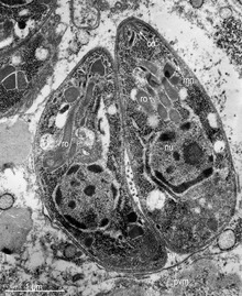
The parasitophorous vacuole is formed during cell invasion, when the parasite uses part of the membrane of the host cell to form a parasitophorous vacuolar membrane (PVM). The PVM surrounds the intracellular parasite, creating a separate bubble of cytoplasm-filled plasma membrane within the host cell. The rhoptry and the microneme, special secretory organelles found in apicomplexan parasites, play a major role in the formation of the vacuole.[2][3] The PVM is extensively re-modelled by parasitic proteins.[4] One theory is that the microneme works with the rhoptry and the rhoptry secretes proteins to create the PVM, while the microneme binds to the surface of red blood cells, allowing the parasite to more easily enter into the cell.[5]
The PV is not a true vacuole, but resembles one under the microscope.[5]
References
- ^ a b Laliberté, J.; Carruthers, V. B. (2008). "Host cell manipulation by the human pathogen Toxoplasma gondii". Cellular and Molecular Life Sciences. 65 (12): 1900–1915. doi:10.1007/s00018-008-7556-x. ISSN 1420-682X. PMC 2662853. PMID 18327664.
- ^ Kemp LE, Yamamoto M, Soldati-Favre D (2013). "Subversion of host cellular functions by the apicomplexan parasites". FEMS Microbiol. Rev. 37 (4): 607–31. doi:10.1111/1574-6976.12013. PMID 23186105.
- ^ Richard, D; et al. (March 2009). "Identification of rhoptry trafficking determinants and evidence for a novel sorting mechanism in the malaria parasite Plasmodium falciparum". PLOS Pathogens. 5 (3): e1000328. doi:10.1371/journal.ppat.1000328. PMC 2648313. PMID 19266084.
- ^ Burda, Paul-Christian; et al. (28 February 2018). "BioID Reveals Novel Proteins of the Plasmodium Parasitophorous Vacuole Membrane". mSphere. 3 (1): e00522–17. doi:10.1128/mSphere.00522-17. PMC 5784244. PMID 29404413.
- ^ a b Lingelbach K, Joiner KA (1998). "The parasitophorous vacuole membrane surrounding Plasmodium and Toxoplasma: an unusual compartment in infected cells". J. Cell Sci. 111 (11): 1467–75. doi:10.1242/jcs.111.11.1467. PMID 9580555.
This microbiology-related article is a stub. You can help Wikipedia by expanding it. |