Muscular system
This article needs more reliable medical references for verification or relies too heavily on primary sources. (October 2016) |  |
| Muscular system | |
|---|---|
 The human muscles, seen from the front, 19th century illustration | |
| Details | |
| Identifiers | |
| Latin | systema musculare |
| TA98 | A04.0.00.000 A04.6.02.001 A04.7.02.001 |
| TA2 | 1975 |
| FMA | 72954 |
| Anatomical terminology | |
The muscular system is an organ system consisting of skeletal, smooth, and cardiac muscle. It permits movement of the body, maintains posture, and circulates blood throughout the body.[1] The muscular systems in vertebrates are controlled through the nervous system although some muscles (such as the cardiac muscle) can be completely autonomous. Together with the skeletal system in the human, it forms the musculoskeletal system, which is responsible for the movement of the body.[2]
Types

There are three distinct types of muscle: skeletal muscle, cardiac or heart muscle, and smooth (non-striated) muscle. Muscles provide strength, balance, posture, movement, and heat for the body to keep warm.[3]
There are approximately 640 muscles in an adult male human body.[4] A kind of elastic tissue makes up each muscle, which consists of thousands, or tens of thousands, of small muscle fibers. Each fiber comprises many tiny strands called fibrils, impulses from nerve cells control the contraction of each muscle fiber.
Skeletal
Skeletal muscle, is a type of striated muscle, composed of muscle cells, called muscle fibers, which are in turn composed of myofibrils. Myofibrils are composed of sarcomeres, the basic building blocks of striated muscle tissue. Upon stimulation by an action potential, skeletal muscles perform a coordinated contraction by shortening each sarcomere. The best proposed model for understanding contraction is the sliding filament model of muscle contraction. Within the sarcomere, actin and myosin fibers overlap in a contractile motion towards each other. Myosin filaments have club-shaped myosin heads that project toward the actin filaments,[1][3][5] and provide attachment points on binding sites for the actin filaments. The myosin heads move in a coordinated style; they swivel toward the center of the sarcomere, detach and then reattach to the nearest active site of the actin filament. This is called a ratchet type drive system.[5]
This process consumes large amounts of adenosine triphosphate (ATP), the energy source of the cell. ATP binds to the cross-bridges between myosin heads and actin filaments. The release of energy powers the swiveling of the myosin head. When ATP is used, it becomes adenosine diphosphate (ADP), and since muscles store little ATP, they must continuously replace the discharged ADP with ATP. Muscle tissue also contains a stored supply of a fast-acting recharge chemical, creatine phosphate, which when necessary can assist with the rapid regeneration of ADP into ATP.[6]
Calcium ions are required for each cycle of the sarcomere. Calcium is released from the sarcoplasmic reticulum into the sarcomere when a muscle is stimulated to contract. This calcium uncovers the actin-binding sites. When the muscle no longer needs to contract, the calcium ions are pumped from the sarcomere and back into storage in the sarcoplasmic reticulum.[5]
There are approximately 639 skeletal muscles in the human body.
-
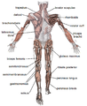
 Skeletal muscles, viewed from the front
 -
 Skeletal muscles, viewed from the back


Cardiac
Heart muscle is striated muscle but is distinct from skeletal muscle because the muscle fibers are laterally connected. Furthermore, just as with smooth muscles, their movement is involuntary. Heart muscle is controlled by the sinus node influenced by the autonomic nervous system.[1][3]
Smooth
Smooth muscle contraction is regulated by the autonomic nervous system, hormones, and local chemical signals, allowing for gradual and sustained contractions. This type of muscle tissue is also capable of adapting to different levels of stretch and tension, which is important for maintaining proper blood flow and the movement of materials through the digestive system.
Physiology
Contraction
Neuromuscular junctions are the focal point where a motor neuron attaches to a muscle. Acetylcholine, (a neurotransmitter used in skeletal muscle contraction) is released from the axon terminal of the nerve cell when an action potential reaches the microscopic junction called a synapse. A group of chemical messengers across the synapse and stimulate the formation of electrical changes, which are produced in the muscle cell when the acetylcholine binds to receptors on its surface. Calcium is released from its storage area in the cell's sarcoplasmic reticulum. An impulse from a nerve cell causes calcium release and brings about a single, short muscle contraction called a muscle twitch. If there is a problem at the neuromuscular junction, a very prolonged contraction may occur, such as the muscle contractions that result from tetanus. Also, a loss of function at the junction can produce paralysis.[5]
Skeletal muscles are organized into hundreds of motor units, each of which involves a motor neuron, attached by a series of thin finger-like structures called axon terminals. These attach to and control discrete bundles of muscle fibers. A coordinated and fine-tuned response to a specific circumstance will involve controlling the precise number of motor units used. While individual muscle units contract as a unit, the entire muscle can contract on a predetermined basis due to the structure of the motor unit. Motor unit coordination, balance, and control frequently come under the direction of the cerebellum of the brain. This allows for complex muscular coordination with little conscious effort, such as when one drives a car without thinking about the process.[5][7]
Tendon
A tendon is a piece of connective tissue that connects a muscle to a bone.[8] When a muscle intercept, it pulls against the skeleton to create movement. A tendon connects this muscle to a bone, making this function possible.
Aerobic and anaerobic muscle activity
At rest, the body produces the majority of its ATP aerobically in the mitochondria[9] without producing lactic acid or other fatiguing byproducts. During exercise, the method of ATP production varies depending on the fitness of the individual as well as the duration and intensity of exercise. At lower activity levels, when exercise continues for a long duration (several minutes or longer), energy is produced aerobically by combining oxygen with carbohydrates and fats stored in the body.[6][10]
During activity that is higher in intensity, with possible duration decreasing as intensity increases, ATP production can switch to anaerobic pathways, such as the use of the creatine phosphate and the phosphagen system or anaerobic glycolysis. Aerobic ATP production is biochemically much slower and can only be used for long-duration, low-intensity exercise, but produces no fatiguing waste products that can not be removed immediately from the sarcomere and the body, and it results in a much greater number of ATP molecules per fat or carbohydrate molecule. Aerobic training allows the oxygen delivery system to be more efficient, allowing aerobic metabolism to begin quicker. Anaerobic ATP production produces ATP much faster and allows near-maximal intensity exercise, but also produces significant amounts of lactic acid which render high-intensity exercise unsustainable for more than several minutes. The phosphagen system is also anaerobic. It allows for the highest levels of exercise intensity, but intramuscular stores of phosphocreatine are very limited and can only provide energy for exercises lasting up to ten seconds. Recovery is very quick, with full creatine stores regenerated within five minutes.[6][11]
Clinical significance
This section needs expansion. You can help by adding to it. (November 2017) |
Multiple diseases can affect the muscular system.
Muscular Dystrophy

Muscular dystrophy is a group of disorders associated with progressive muscle weakness and loss of muscle mass. These disorders are caused by mutations in a person’s genes.[12] The disease affects between 19.8 and 25.1 per 100,000 person-years globally.[13]
There are more than 30 types of muscular dystrophy. Depending on the type, muscular dystrophy can affect the patient's heart and lungs, and/or their ability to move, walk, and perform daily activities. The most common types include:
- Duchenne muscular dystrophy (DMD) and Becker muscular dystrophy (BMD)
- Myotonic dystrophy
- Limb-Girdle (LGMD)
- Facioscapulohumeral dystrophy (FSHD)
- Congenital dystrophy (CMD)
- Distal (DD)
- Oculopharyngeal dystrophy (OPMD)
- Emery-Dreifuss (EDMD)
See also
References
- ^ a b c Ross MH, Wojciech P (2011). Histology: a text and atlas: with correlated cell and molecular biology (6th ed.). Philadelphia: Wolters Kluwer/Lippincott Williams & Wilkins Health. ISBN 9780781772006. OCLC 548651322.
- ^ Standring S, Gray H (2016). Gray's anatomy : the anatomical basis of clinical practice (Forty-first ed.). [Philadelphia]. ISBN 9780702052309. OCLC 920806541.
{{cite book}}: CS1 maint: location missing publisher (link) - ^ a b c Mescher AL, Junqueira LC (2013-02-22). Junqueira's basic histology : text and atlas (Thirteenth ed.). New York. ISBN 9780071807203. OCLC 854567882.
{{cite book}}: CS1 maint: location missing publisher (link) - ^ "THE MUSCULAR SYSTEM" (PDF). www.uc.edu.
- ^ a b c d e Hall JE, Guyton AC (2011). Guyton and Hall textbook of medical physiology (Twelfth ed.). Philadelphia, Pa. ISBN 9781416045748. OCLC 434319356.
{{cite book}}: CS1 maint: location missing publisher (link) - ^ a b c Lieberman M, Peet A (2018). Marks' basic medical biochemistry : a clinical approach (Fifth ed.). Philadelphia. ISBN 9781496324818. OCLC 981908072.
{{cite book}}: CS1 maint: location missing publisher (link) - ^ Blumenfeld H (2010). Neuroanatomy through clinical cases (2nd ed.). Sunderland, Mass.: Sinauer Associates. ISBN 9780878930586. OCLC 473478856.
- ^ "Tendon vs. ligament: MedlinePlus Medical Encyclopedia Image". medlineplus.gov.
- ^ Abercrombie M, Hickman CJ, Johnson ML (1973). A Dictionary of Biology. Penguin reference books (6th ed.). Middlesex (England), Baltimore (U.S.A.), Ringwood (Australia): Penguin Books. p. 179. OCLC 943860.
- ^ Scott C (December 2005). "Misconceptions about Aerobic and Anaerobic Energy Expenditure". Journal of the International Society of Sports Nutrition. 2 (2): 32–37. doi:10.1186/1550-2783-2-2-32. PMC 2129144. PMID 18500953.
- ^ Spriet LL (January 1992). "Anaerobic metabolism in human skeletal muscle during short-term, intense activity". Canadian Journal of Physiology and Pharmacology. 70 (1): 157–165. doi:10.1139/y92-023. PMID 1581850.
- ^ CDC (2022-11-21). "What is Muscular Dystrophy? | CDC". Centers for Disease Control and Prevention. Retrieved 2023-05-05.
- ^ Theadom A, Rodrigues M, Roxburgh R, Balalla S, Higgins C, Bhattacharjee R, Jones K, Krishnamurthi R, Feigin V (2014-12-16). "Prevalence of Muscular Dystrophies: A Systematic Literature Review". Neuroepidemiology. 43 (3–4): 259–268. doi:10.1159/000369343. hdl:10292/13206. ISSN 0251-5350. PMID 25532075. S2CID 2426923.
Further reading
- Cartee GD, Hepple RT, Bamman MM, Zierath JR (June 2016). "Exercise Promotes Healthy Aging of Skeletal Muscle". Cell Metabolism. 23 (6): 1034–1047. doi:10.1016/j.cmet.2016.05.007. PMC 5045036. PMID 27304505.
- Murphy AC, Muldoon SF, Baker D, Lastowka A, Bennett B, Yang M, Bassett DS (January 2018). "Structure, function, and control of the human musculoskeletal network". PLOS Biology. 16 (1): e2002811. doi:10.1371/journal.pbio.2002811. PMC 5773011. PMID 29346370.
External links
- "Muscle". Cleveland Clinic.
- Online Muscle Tutorial
- GetBody Smart Muscle system tutorials and quizzes
- MedBio.info Archived 2011-02-05 at the Wayback Machine Use and formation of ATP in muscle
- CS1 maint: location missing publisher
- Articles with short description
- Short description is different from Wikidata
- Articles needing additional medical references from October 2016
- All articles needing additional references
- Articles requiring reliable medical sources
- Articles needing additional references from October 2016
- Articles to be expanded from November 2017
- All articles to be expanded
- Articles using small message boxes
- Webarchive template wayback links
- Articles with GND identifiers
- Articles with J9U identifiers
- Articles with LCCN identifiers
- Articles with NKC identifiers
- Articles with TA98 identifiers
- Muscular system