Milium (dermatology)
| Milia | |
|---|---|
| Other names: Milk spot, oil seed[citation needed] | |
.jpg) | |
| Milia | |
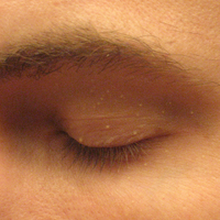
| Symptoms | Single or multiple small 1-2mm pearly-white bumps usually on face and and around eyes[1] |
| Usual onset | Any age[2] |
| Types | Neonatal, primary, juvenile, milia en plaque, traumatic, drug-induced[1] |
| Causes | Congenital, trauma, topical corticosteroids, dermabrasion, associated with DLE[2] |
| Diagnostic method | Visualisation, biopsy[1] |
| Differential diagnosis | Erythropoietic protoporphyria, mucinoses, lipoid proteinosis, adnexal tumors, primary systemic amyloidosis (adults)[3] |
| Treatment | Usually none,[1] laser therapy, dermabrasion, intense pulsed light, chemical peels, cryotherapy[3] |
| Frequency | Common[1] |
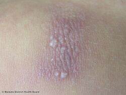
A milium (plural milia), is a small 1-2mm white bump in the skin of typically the face and around the eyes, which can appear on its own or more usually as several.[2] Types include neonatal, primary, juvenile, milia en plaque, traumatic, and drug-induced.[1] They are sometimes on the genitalia, often mistaken by those affected as warts or other sexually transmitted diseases.[citation needed] Milia can also be confused with stubborn whiteheads.[citation needed]
They can be congenital, triggered by trauma, topical corticosteroids or dermabrasion, or associated with discoid lupus erythematosus.[2] It is a clog of the eccrine sweat gland. It is a keratin-filled cyst that can appear just under the epidermis or on the roof of the mouth.[4][5]: 780
Milia are commonly associated with newborn babies but can appear on people of all ages.[6]: 680
In children, milia often but not always disappear within two to four weeks. For adults, they can be removed by a physician (a dermatologist will have specialist knowledge in this area). A common method that a dermatologist will use to remove a milium is to nick the skin with a #11 surgical blade and then use a comedone extractor to press the cyst out.[7][clarification needed]
-
Milia
-
Milia
-

Milia
.jpg)


See also
Epidemiology

References
- ↑ 1.0 1.1 1.2 1.3 1.4 1.5 "Milium". dermnetnz.org. Archived from the original on 15 August 2021. Retrieved 25 September 2021.
- ↑ 2.0 2.1 2.2 2.3 Johnstone, Ronald B. (2017). "16. Cysts, sinuses and pits". Weedon's Skin Pathology Essentials (2nd ed.). Elsevier. p. 338. ISBN 978-0-7020-6830-0. Archived from the original on 2021-05-25. Retrieved 2021-09-25.
- ↑ 3.0 3.1 Quist, Jennifer; Quist, Sven; Gollnick, Harold (2012). "48. Deposition diseases". In Bolognia, Jean L.; Jorizzo, Joseph L.; Schaffer, Julie V. (eds.). Dermatology. Vol. 1. Elsevier Health Sciences. pp. 712–714. ISBN 978-0-7234-3571-6. Archived from the original on 2021-09-25. Retrieved 2021-09-25.
- ↑ "milium" at Dorland's Medical Dictionary
- ↑ Freedberg, et al. (2003). Fitzpatrick's Dermatology in General Medicine. (6th ed.). McGraw-Hill. ISBN 0-07-138076-0.
- ↑ James, William D.; Berger, Timothy G.; et al. (2006). Andrews' Diseases of the Skin: Clinical Dermatology. Saunders Elsevier. ISBN 0-7216-2921-0.
- ↑ Burnett, Mark E.; Levitt, Jacob O. (2015). "Incision and Drainage (Abscesses, Acne, and Milia)". In Levitt, J.; Sobak, J. (eds.). Safety in Office-Based Dermatologic Surgery. Switzerland: Spring, Cham. pp. 119–128. doi:10.1007/978-3-319-13347-8_13.
| Classification | |
|---|---|
| External resources |