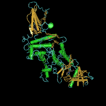
Juvenile-hormone esterase
| juvenile hormone esterase | |||||||||
|---|---|---|---|---|---|---|---|---|---|
 | |||||||||
| Identifiers | |||||||||
| EC no. | 3.1.1.59 | ||||||||
| CAS no. | 50812-15-2 | ||||||||
| Databases | |||||||||
| IntEnz | IntEnz view | ||||||||
| BRENDA | BRENDA entry | ||||||||
| ExPASy | NiceZyme view | ||||||||
| KEGG | KEGG entry | ||||||||
| MetaCyc | metabolic pathway | ||||||||
| PRIAM | profile | ||||||||
| PDB structures | RCSB PDB PDBe PDBsum | ||||||||
| Gene Ontology | AmiGO / QuickGO | ||||||||
| |||||||||
The enzyme juvenile hormone esterase (EC 3.1.1.59, systematic name methyl-(2E,6E,10R)-10,11-epoxy-3,7,11-trimethyltrideca-2,6-dienoate acylhydrolase, JH esterase) catalyzes the hydrolysis of juvenile hormone:
- (1) juvenile hormone I + H2O = juvenile hormone I acid + methanol
- (2) juvenile hormone III + H2O = juvenile hormone III acid + methanol
Nomenclature and function
This enzyme belongs to the family of hydrolases, specifically those acting on carboxylic ester bonds. The systematic name of this enzyme class is methyl-(2E,6E)-(10R,11S)-10,11-epoxy-3,7,11-trimethyltrideca-2,6-dienoate acylhydrolase. Other names in common use include JH esterase, juvenile hormone esterase, and juvenile hormone carboxyesterase.
Juvenile hormone (JH) controls insect metamorphosis.[1] High JH titers maintain the larval state while a decrease in the JH titer initiates the pupation sequence [2] as well as a change in tissue commitment away from synthesis of larval tissues to pupal tissues at the pupal stage.[3] The drop in JH titer at the beginning of the last larval instar in the Lepidoptera appears to be due to a combination of increased metabolism [4] and decreased synthesis.[5] In the Lepidoptera, JH is initially metabolized by ester hydrolysis;[6] esterases capable of hydrolyzing JH are detectable in the hemolymph at times during the last larval instar that appear to coincide with reported drops in the JH titre.[7] The JHE's are also selective for the 2E methyl ester of the naturally occurring JH's.[8] These studies suggest that the JHE's may be important in the regulation of the JH titre and therefore involved in the initiation of and the commitment to the pupal stage. JHE's appear to be produced by the fat body[9] and this production can be stimulated by exogenous JH in Hyalophora pupae, a stage devoid of JHE activity.[10] Stimulation of JHE activity by JH has also been noted recently in adults of Leptinotarsa decemlineata [11] and pupae of Galleria mellonella.[12] However, to date, no reported studies have examined this phenomenon during the last larval instar when these enzymes are thought to be of primary importance. Thus this laboratory undertook an investigation of the hemolymph JHE regulation during the last larval instar of the cabbage looper, Trichoplusia ni.
JH esterase induction
Juvenile hormone esterase is induced by factors naturally occurring in the head of insects.[13] In addition, it is induced by treatment of insects with either natural juvenile hormone, with JH I being the most potent inducer.[14] Synthetic agonists of JH have been shown to possess this same activity, albeit at a lower potency than JH I.[15] In another study, it has been shown that factors present in the head of the insect are potent inducers of JH activity.[16] Starvation of lepidopteran larvae also induces appearance of JH esterase.[17]
JH esterase inhibitors
A number of compounds have been discovered which are potent inhibitors of JH esterase. Many of these are insecticides falling into two major structural groups, the phosphoamidothiolates and S-phenylphosphates; carbamate insecticides were also tested.[18] By far the most potent inhibitor was an ethoxythiophenylphospamidothiolate, with IC50 < 1 nM. Of particular interest in this study is that ethyl and isopropyl analogs of natural JHs were NOT cleaved by the esterase, showing that it is methyl ester specific. JH I and JH III were tested at nominal concentrations of 5 μM. Later a trifluoromethyl ketone (3-octylthio-1,1,1-trifluoro-2-propanone) was shown to be a highly potent, high affinity slow, tight binding inhibitor of the JH esterase of Trichoplusia ni, the same Lepidopteran which was used in the other study in this section.[19] This study reported very sophisticated kinetic analyses of the inhibition of this compound (acronym OTFP), JH I was shown to be degraded more readily by the enzyme than JH III, with a Km value about twice the value of JH III.
JH esterase fluctuations with time and relation to insect development
JH esterase and JH epoxide hydrolase are crucial in terminating the action of JH. The role of juvenile hormone binding proteins are also important, as they afford juvenile hormone protection from hydrolytic enzymes.[20] This makes for a very complicated scenario that is difficult to investigate, and also difficult to distinguish between different species. Very detailed studies have been done on JH, JH acid, ecdysone, and JH titers have been done in precisely timed larvae of Manduca sexta as a function of development during the fifth larval stadium. In these larvae the principle JH are JH I, and JH II, with low levels of JH 0 and JH III. There is a large peak of JH I and II at the end of the fourth stadium, accompanied by lower levels of their acid metabolites. Then a broad peak of JH esterase starts on day 1.5 to day 4. Subsequently, ecdysteroid titers rise slightly on day 3.5, then a massive peak of ecdysteroid starting on day 5 persisting at somewhat lower levels to day 5. This is accompanied by a sharp peak of JH I and JH II, beginning on day 4 and ending on day 6. JH I acid titers are almost the same as JH I titer, except on day 7 when is a sharp peak of only JH I acid. This is just as ecdysteroid titers are decreasing.[21] A very similar timing of peaks of JH esterase and ecdysone has been observed in Galleria mellonella.[22] These data are consistent with a classical model for lepidopterans where JH is high at each larval molt, but must rise together with ecdysone prior to pupation to initiate the pupal molt. They are also consistent with a model advanced by others that corpora allata maintained in vitro of day 0 M. sexta larva secrete high levels of JH, but that the shift to producing only JH acid at day 4, which is then methylated by imaginal discs to generate the JH peak.[23] However, secretion of the relative amounts of JH produced by CA of Manduca sexta has been found to differ considerably from in vivo titers.[24] Investigation of JH titers in Trichoplusia ni have led to very similar conclusions as regards the timing of pulses of JH temporally and with respect to edysone secretion. However, the principle JH in this species is JH II. Injection of an esterase inhibitor, EPPAT, was found to increase juvenile hormone titers, and starvation was found to increase juvenile hormone titers. In addition, parasitization of larvae with Chelonus sp. (Hymenoptera) was found to decrease JH II titers, but to cause an increase in JH III titers, apparently derived from the parasite.[25]
JH esterase protein structure
The crystal structure of esterase from the tobacco hornworm Manduca sexta has been solved in complex with the transition state analogue inhibitor 3-octylthio-1,1,1-trifluoropropan-2-one (OTFP) covalently bound to the active site. This crystal structure contains a long, hydrophobic binding pocket with the solvent-inaccessible catalytic triad located at the end. The structure explains many of the interactions observed between JHE and its substrates and inhibitors, such as the preference for methyl esters vs. ethyl or isopropyl esters, and long hydrophobic backbones.[26] The enzyme is extremely efficient, with a cat/Km of at least 3 x 10⁷ M-1 s-1. The primary sequence is 583 amino acids long, with a 22 amino acid protein. The calculated Mr of the active form is 62.1 kDa.
Written References
Abdel-Aal, Y.A.I., Roe, R., Hammock, B.D., 1984. Kinetic properties of the inhibition juvenile hormone esterase by two trifluoromethylketones and O-ethyl, S-phenyl phosphoramidothioate. Pest. Biochem. Physiol. 21, 232-241.
Baker, F.C., Tsai, L.W., Reuter, C.C., Schooley, D.A., 1987. In vivo fluctuation of JH, JH acid, and ecdysteroid titer, and JH esterase activity, during development of fifth stadium Manduca sexta. Insect Biochem. 17, 989-996.
Braun, R.P., Wyatt, G.R., 1995. Growth of the male accessory gland in adult locusts: Roles of juvenile hormone, JH esterase, and JH binding proteins. Arch.Insect Biochem.Physiol. 30, 383-400.
De Kort, C.A.D., Granger, N.A., 1996. Regulation of JH titers: The relevance of degradative enzymes and binding proteins. Arch. Insect Biochem. Physiol. 33, 1-26.
Gilbert, L.I., Goodman, W., Bollenbacher, W.E., 1977. Biochemistry of regulatory lipids and sterols in insects., in: Goodwin, T.W. (Ed.), Biochemistry of Lipids II. International Review of Biochemistry. University Park Press, Baltimore, pp. I-50.
Hammock, B., Nowock, J., Goodman, W., Stamoudis, V., Gilbert, L.I., 1975. Influence of Hemolymph-Binding Protein on Juvenile-Hormone Stability and Distribution in Manduca Sexta Fat-Body and Imaginal Disks In vitro. Molecular and Cellular Endocrinology 3, 167-184.
Hammock, B.D., Quistad, G.B., 1976. The degradative metabolism of juvenoids by insects, in: Gilbert, L.I. (Ed.), The Juvenile Hormones. Plenum Press, New York, pp. 374–393.
Hammock, B.D., Sparks, T.C., Mumby, S.M., 1977. Selective inhibition of JH esterases from cockroach hemolymph. Pest. Biochem. Physiol. 7, 517-530.
Hwanghsu, K., Reddy, G., Kumaran, A.K., Bollenbacher, W.E., Gilbert, L.I., 1979. Correlations between Juvenile Hormone Esterase-Activity, Ecdysone Titer and Cellular Reprogramming in Galleria Mellonella. J. Insect Physiol. 25, 105-111.
Jones, G., Hanzlik, T., Hammock, B.D., Schooley, D.A., Miller, C.A., Tsai, L.W., Baker, F.C., 1990. The Juvenile Hormone Titre During the Penultimate and Ultimate Larval Stadia of Trichoplusia ni. J. Insect Physiol. 36, 77-83.
Jones, G., Wing, K.D., Jones, D., Hammock, B.D., 1980. The source and action of head factors regulating juvenile hormone esterase in larvae of the cabbage looper, Trichoplusia ni. J. Insect Physiol. 27, 85-91.
Kramer, S.J., 1978. Regulation of the activity of JH-specific esterases in the Colorado potato beetle, Leptinotarsa decemlineata. J. Insect Physiol. 24, 743-747.
Nijhout, H., Williams, C., 1974. Control of Moulting and Metamorphosis in the Tobacco Hornworm, Manduca Sexta (L.): Cessation of Juvenile Hormone Secretion as a Trigger for Pupation J. Exp. Biol. 61, 493-450.
Nijhout, H.F., 1975. Dynamics of juvenile hormone action in larvae of the tobacco horn worm. Biological Bulletin of the Marine Biological Laboratory, Woods Hole 149, 568-579.
Nowock, J., GILBERT, L., 1976a. In vitro analysis of factors regulating the juvenile hormone titer of insects, in: Kurstack, E., Maramorosch, K. (Eds.), Invertebrate Tissue Culture. Academic Press, New York, pp. 203–212.
Nowock, J., Gilbert, L.I., 1976b. In vitro analysis of factors regulating the juvenile hormone titer of insects, in: K., K.E.a.M. (Ed.), Invertebrate Tissue Culture. Academic Press, New York, pp. 203–212.
Plapp, F.W., Jr., Cariño, F.A., Wei, V.K., 1998. A juvenile hormone binding protein from the house fly and its possible relationship to insecticide resistance. Arch. Insect Biochem. Physiol. 37, 64-72.
Prestwich, G.D., Wojtasek, H., Lentz, A.J., Rabinovich, J.M., 1996. Biochemistry of proteins that bind and metabolize juvenile hormones. Arch. Insect Biochem. Physiol. 32, 407-419.
Reddy, G., Hwanghsu, K., Kumaran, A.K., 1979. Factors Influencing Juvenile Hormone Esterase-Activity in the Wax Moth, Galleria-Mellonella. J. Insect Physiol. 25, 65-71.
Riddiford, L.M., 1976. Hormonal control of insect epidermal cell commitment in vitro. Nature 259, 115-117.
Sanburg, L.L., Kramer, K.J., Kezdy, F.J., Law, J.H., 1975a. Juvenile hormone-specific esterases in the haemolymph of the tobacco hornworm, Manduca sexta. J. Insect Physiol. 21, 873-887.
Sanburg, L.L., Kramer, K.J., Kezdy, F.J., Law, J.H., Oberlander, H., 1975b. Role of juvenile hormone esterases and carrier proteins in insect development. Nature 253, 266-267.
Slade, M., Zibitt, C.H., 1972. Metabolism of Cecropia Juvenile Hormone in Insects and in Mammals, in: Menn, J.J., Beroza, M. (Eds.), Insect Juvenile Hormones: Chemistry and Action. Academic Press, New York, pp. 155–176.
Sparagana, S.P., Bhaskaran, G., Barrera, P., 1985. Juvenile hormone acid methyltransferase activity in imaginal discs of Manduca sexta prepupae. Arch. Insect Biochem. Physiol. 2, 191-202.
Sparagana, S.P., Bhaskaran, G., Dahm, K.H., Riddle, V., 1984. Juvenile hormone production, juvenile hormone esterase, and juvenile hormone acid methyltransferase in corpora allata of Manduca sexta. J. Exp. Zool. 230, 309-313.
Sparks, T.C., Hammock, B.D., 1979. Induction and regulation of juvenile hormone esterases during the last larval instar of the cabbage looper, Trichoplusia ni. J. Insect Physiol. 25, 551-560.
Sparks, T.C., Hammock, B.D., Riddiford, L.M., 1983. The haemolyph juvenile hormone esterase of Manduca sexta (L.)-inhibition and regulation. Insect Biochem. 13, 529-541.
Sparks, T.C., Wing, K.D., Hammock, B.D., 1979. Effects of the anti hormone-hormone mimic ETB on the induction of insect juvenile hormone esterase in Trichoplusia ni. Life Sci. 25, 445-450.
Vince, R.K., Gilbert, L.I., 1977. Juvenile hormone esterase activity in precisely timed last instar larvae and pharate pupae of Manduca sexta. Insect Biochem. 7, 115-120.
Weirich, G., Wren, J., 1973. The substrate specificity of juvenile hormone esterase from Manduca sexta haemolymph. Life Sci. 13, 213-226.
Weirich, G.F., Wren, J., 1976. Juvenile-hormone esterase in insect development: a comparative study. Physiological Zoology 49, 341-350.
Whitmore, D., Gilbert, L.I., Ittycher.Pi, 1974. Origin of Hemolymph Carboxylesterases Induced by Insect Juvenile-Hormone. Molecular and Cellular Endocrinology 1, 37-54.
Wogulis, M., Wheelock, C.E., Kamita, S.G., Hinton, A.C., Whetstone, P.A., Hammock, B.D., Wilson, D.K., 2006. Structural Studies of a Potent Insect Maturation Inhibitor Bound to the Juvenile Hormone Esterase of Manduca sexta. Biochemistry 45, 4045-4057.
Further reading
- Foucher AL, McIntosh A, Douce G, Wastling J, Tait A, Turner CM (2006). "A proteomic analysis of arsenical drug resistance in Trypanosoma brucei". Proteomics. 6 (9): 2726–32. doi:10.1002/pmic.200500419. PMID 16526094.
- Mitsui T, Riddiford LM, Bellamy G (1979). "Metabolism of juvenile hormone by the epidermis of the tobacco hornworm (Manduca sexta)". Insect Biochem. 9 (6): 637–643. doi:10.1016/0020-1790(79)90103-3.
References
- ^ Gilbert et al., 1977
- ^ Nijhout and Williams, 1974
- ^ Riddiford, 1976
- ^ Nowock and GILBERT, 1976a; Sanburg et al., 1975a; Sanburg et al., 1975b
- ^ Nijhout, 1975
- ^ Hammock and Quistad, 1976; Slade and Zibitt, 1972
- ^ Vince and Gilbert, 1977:Sparks, 1979 #1170; Weirich and Wren, 1973
- ^ Hammock et al., 1977; Weirich and Wren, 1973; Weirich and Wren, 1976
- ^ Hammock et al., 1975; Nowock and Gilbert, 1976b; Whitmore et al., 1974
- ^ Whitmore, 1972 #1160;Whitmore et al., 1974
- ^ Kramer, 1978
- ^ Reddy et al., 1979
- ^ Jones et al., 1980
- ^ Sparks and Hammock, 1979
- ^ Sparks et al., 1979
- ^ Jones et al., 1980
- ^ Sparks et al., 1983
- ^ Hammock et al., 1977
- ^ Abdel-Aal et al., 1984
- ^ Braun and Wyatt, 1995; De Kort and Granger, 1996; Plapp et al., 1998; Prestwich et al., 1996
- ^ Baker et al., 1987
- ^ Hwanghsu et al., 1979
- ^ Sparagana et al., 1985; Sparagana et al., 1984
- ^ Baker et al., 1987
- ^ Jones et al., 1990
- ^ Wogulis et al., 2006