Janeway lesion
| Janeway lesion | |
|---|---|
 | |
| Specialty | |
| Symptoms | Painless red flat marks on palms and soles[1] |
| Usual onset | Sudden |
| Duration | Days to weeks[1] |
| Causes | Infective endocarditis[1] |
| Differential diagnosis | Osler nodes[1] |
Janeway lesions are irregular reddish spots on the palms or soles, seen in infective endocarditis.[1] Unlike Osler nodes, they do not hurt.[1] They may be small bumps or larger bumps but typically only a few millimeters in diameter and can last days to weeks.[1]
Janeway lesions were named by Emanuel Libman for Edward Janeway, a contemporary of Sir William Osler.[2][3]
Definition
Janeway lesions are painless, frequently haemorrhagic lesions seen most commonly on the palms and soles, particularly on the base of the thumb and little finger, and seen in infective endocarditis.[4]
Differential
Osler nodes and Janeway lesions are similar and point to the same diagnostic conclusion. The most significant difference between the two is that Osler nodes present with tenderness, while Janeway lesions do not.[5] Osler nodes are thought to be due to immunologic phenomenon where deposition of immune complexes provoke inflammatory response, leading to swelling, redness and pain. On the contrary, Janeway lesions are thought to be due to embolic phenomenon in cutaneous blood vessels of palms and soles which does not cause pain or least pain. [6]
Pathophysiology
Pathologically, the lesion is described to be a microabscess of the dermis with marked necrosis and inflammatory infiltrate not involving the epidermis.[5]
They are caused by septic emboli which deposit bacteria, forming microabscesses.[7] Organisms may be cultured from the lesions.[8]
Diagnosis
Janeway lesions present as red, painless macules and papules on the palms and soles.[4]
They are not common and are frequently indistinguishable from Osler nodes. Rarely, they have been reported in cases of Systemic lupus erythematosis (SLE), Gonococcemia (disseminated gonorrhoea), haemolytic anaemia and typhoid fever.[4]
They may last days to weeks before completely resolving.[4]
History
Janeway lesions are named for Edward Janeway (1841–1911), a prominent American physician, pathologist and contemporary of Sir William Osler, who initially described "peculiar skin lesions" in some people with endocarditis, in a paper published in 1899.[2] The term was first used by internist and pathologist Emanuel Libman, who reported the lesions in his paper of 1906 and explained his reasoning for using the term "Janeway lesions" in a footnote in 1923. Osler never mentioned Janeway lesions. The inclusion into Osler's 1925 textbook came six years after Osler died.[2]
Additional images
-
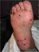
Multiple dark maculae on the sole of the right foot resembling septic emboli/Janeway lesions
-
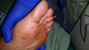
Janeway lesions (arrows) on the toes and sole, seen in a patient with massive aortic valve vegetation.


References
- ↑ 1.0 1.1 1.2 1.3 1.4 1.5 1.6 Parashar, Krishan; Daveluy, Steven (2022). "Osler Node and Janeway Lesions". StatPearls. StatPearls Publishing. PMID 32491553. Archived from the original on 2022-05-16. Retrieved 2022-11-04.
- ↑ 2.0 2.1 2.2 Jordan Prutkin, W. Bruce Fye (2006). "Edward G. Janeway, Clinician and Pathologist". Clinical Cardiology. 29 (8): 376–377. doi:10.1002/clc.4960290815. PMC 6654287. PMID 16933584.
{{cite journal}}: CS1 maint: uses authors parameter (link) - ↑ Yale, Steven H.; Tekiner, Halil; Mazza, Joseph J.; Yale, Eileen S.; Yale, Ryan C. (2021). "3. Endocarditis". Cardiovascular Eponymic Signs: Diagnostic Skills Applied During the Physical Examination. Switzerland: Springer. pp. 76–77. ISBN 978-3-030-67595-0. Archived from the original on 2023-07-01. Retrieved 2023-05-28.
- ↑ 4.0 4.1 4.2 4.3 "Osler nodes and Janeway lesions | DermNet NZ". www.dermnetnz.org. Archived from the original on 8 January 2020. Retrieved 2 October 2019.
- ↑ 5.0 5.1 Farrior, J.B.; Silverman M.E. (1976). "A consideration of the differences between a Janeway's lesion and an Osler's node in infectious endocarditis". Chest. 70 (2): 239–243. doi:10.1378/chest.70.2.239. PMID 947688.
- ↑ "Why Osler's Nodes are Painful while Janeway Lesions are Painless?". YouTube. Archived from the original on 2022-02-12. Retrieved 2022-08-17.
- ↑ Mandell, Douglas, Bennett's Principles and Practice of Infectious Diseases, 7th ed., Churchill Livingstone (2009).
- ↑ Patterson, James W. (2016). "8. The Vasculopathic Reaction Pattern". Weedon's Skin Pathology (4th ed.). Churchill Livingston. pp. 239–240. ISBN 9780702051838. Archived from the original on 2022-11-12. Retrieved 2022-08-17.
External links
| Classification |
|---|