Syringomyelia
| Syringomyelia | |
|---|---|
.png) | |
| An idiopathic syrinx | |
| Pronunciation | |
| Specialty | Neurosurgery |
Syringomyelia is a generic term referring to a disorder in which a cyst or cavity forms within the spinal cord. Often, syringomyelia is used as a generic term before an etiology is determined. [3] This cyst, called a syrinx, can expand and elongate over time, destroying the spinal cord. The damage may result in loss of feeling, paralysis, weakness,[4] and stiffness in the back, shoulders, and extremities. Syringomyelia may also cause a loss of the ability to feel extremes of hot or cold, especially in the hands. It may also lead to a cape-like bilateral loss of pain and temperature sensation along the upper chest and arms. Each patient experiences a different combination of symptoms. These symptoms typically vary depending on the extent and, often more critically, on the location of the syrinx within the spinal cord.
Syringomyelia has a prevalence estimated at 8.4 cases per 100,000 people,[5] with symptoms usually beginning in young adulthood. Signs of the disorder tend to develop slowly, although sudden onset may occur with coughing, straining, or myelopathy.
Signs and symptoms
Syringomyelia causes a wide variety of neuropathic symptoms, due to damage to the spinal cord. Patients may experience severe chronic pain, abnormal sensations and loss of sensation, particularly in the hands. Some patients experience paralysis or paresis, temporarily or permanently. A syrinx may also cause disruptions in the parasympathetic and sympathetic nervous systems, leading to abnormal body temperature or sweating, bowel control issues, or other problems. If the syrinx is higher up in the spinal cord or affecting the brainstem, as in syringobulbia, vocal cord paralysis, ipsilateral tongue wasting, trigeminal nerve sensory loss, and other signs may be present.[6] Rarely, bladder stones can occur at the onset of weakness in the lower extremities.[7] Classically, syringomyelia spares the dorsal column/medial lemniscus of the spinal cord, leaving pressure, vibration, touch and proprioception intact in the upper extremities. Neuropathic arthropathy, also known as a Charcot joint, can occur, particularly in the shoulders, in patients with syringomyelia.[8] The loss of sensory fibers to the joint is theorized to lead to degeneration of the joint over time.[9]
Cause
Generally, there are two forms of syringomyelia: congenital and acquired. Syringomyelia is generally a chronic disorder that occurs over time, resulting in muscular atrophy. Acquired Syringomyelia can be caused by a serious physical trauma to the body such as in a road traffic accident. Syringomyelia can also be classified into communicating and noncommunicating forms. Communicating typically occurs due to lesions on the foramen magnum and noncommunicating occurring due to other spinal cord diseases.[10]
Congenital
The first major form relates to an abnormality of the brain called an Arnold–Chiari malformation or Chiari malformation. This is the most common cause of syringomyelia, where the anatomic abnormality, which may be due to a small posterior fossa, causes the lower part of the cerebellum to protrude from its normal location in the back of the head into the cervical or neck portion of the spinal canal. A syrinx may then develop in the cervical region of the spinal cord. Here, symptoms usually begin between the ages of 25 and 40 and may worsen with straining, called a valsalva maneuver, or any activity that causes cerebrospinal fluid pressure to fluctuate suddenly. Some patients, however, may have long periods of stability. Some patients with this form of the disorder also have hydrocephalus, in which cerebrospinal fluid accumulates in the skull, or a condition called arachnoiditis, in which a covering of the spinal cord—the arachnoid membrane—is inflamed.[11] Some cases of syringomyelia are familial, although this is rare.[12]
Acquired
The second major form of syringomyelia occurs as a complication of trauma, meningitis, hemorrhage, a tumor, or arachnoiditis. Here, the syrinx or cyst develops in a segment of the spinal cord damaged by one of these conditions. The syrinx then starts to expand. This is sometimes referred to as noncommunicating syringomyelia. Symptoms may appear months or even years after the initial injury, starting with pain, weakness, and sensory impairment originating at the site of trauma.[citation needed]
The primary symptom of post-traumatic syringomyelia (often referred to using the abbreviation of PTS)[13] is pain, which may spread upward from the site of injury. Symptoms, such as pain, numbness, weakness, and disruption in temperature sensation, may be limited to one side of the body. Syringomyelia can also adversely affect sweating, sexual function, and, later, bladder and bowel control. A typical cause of PTS would be a car accident or similar trauma involving a whiplash injury.[citation needed]
What can make PTS difficult to diagnose is the fact that symptoms can often first appear long after the actual cause of the syrinx occurred (e.g., a car accident occurring and then the patient first experiencing PTS symptoms such as pain, loss of sensation, and reduced ability on the skin to feel varying degrees of hot and cold a number of months after the car accident).[citation needed]
Pathogenesis
The pathogenesis of syringomyelia is debated. The cerebrospinal fluid also serves to cushion the brain. Excess cerebrospinal fluid in the central canal of the spinal cord is called hydromyelia. This term refers to increased cerebrospinal fluid that is contained within the ependyma of the central canal. When fluid dissects into the surrounding white matter forming a cystic cavity or syrinx, the term syringomyelia is applied. As these conditions coexist in the majority of cases, the term syringohydromyelia is applied. The terms are used interchangeably.[citation needed]
It has been observed that obstruction of the cerebrospinal fluid spaces in the subarachnoid space can result in syrinx formation, and alleviation of the obstruction may improve symptoms. A number of pathological conditions can cause an obstruction of the normal cerebrospinal fluid spaces. These include Chiari malformation, spinal arachnoiditis, scoliosis, spinal vertebrae misalignment, spinal tumors, spina bifida, and others. The reasons that blockage of the cerebrospinal fluid space within the subarachnoid space can result in syrinx formation are not fully understood although a small posterior fossa is one known cause. It is unclear if syrinx fluid originates from bulk movement of cerebrospinal fluid into the spinal cord, from bulk transmural movement of blood fluids through the spinal vasculature into the syrinx, or from a combination of both. Recent work suggests that central nervous system compliance is the underlying problem for the central nervous system, and also that hydrocephalus and syringomyelia have related causes.[citation needed]
Diagnosis
Physicians now use magnetic resonance imaging (MRI) to diagnose syringomyelia. The MRI radiographer takes images of body anatomy, such as the brain and spinal cord, in vivid detail. This test will show the syrinx in the spine or any other conditions, such as the presence of a tumor. MRI is safe, painless, and informative and has greatly improved the diagnosis of syringomyelia.[14][15][16][17][18][19][20][21][22][23][24][25][excessive citations]
The physician may order additional tests to help confirm the diagnosis. One of these is called electromyography (EMG), which show possible lower motor neuron damage.Note this test isn't used diagnostically for injuries to the spine but to nerves and muscles.This would be part of a patients rehab routine. In addition, computed axial tomography (CT) scans of a patient's head may reveal the presence of tumors and other abnormalities such as hydrocephalus.[citation needed]
Like MRI and CT scans, another test, called a myelogram, uses radiographs and requires a contrast medium to be injected into the subarachnoid space. Since the introduction of MRI, this test is rarely necessary to diagnose syringomyelia.[citation needed]
The possible causes are trauma, tumors, and congenital defects. It is most usually observed in the part of the spinal cord corresponding to the neck area. Symptoms are due to spinal cord damage and include pain, decreased sensation of touch, weakness, and loss of muscle tissue. The diagnosis is confirmed with a spinal CT, myelogram or MRI of the spinal cord. The cavity may be reduced by surgical decompression.[citation needed]
Furthermore, evidence also suggests that impact injuries to the thorax area highly correlate with the occurrence of a cervical-located syrinx.[citation needed]
Treatment
Surgery
The first step after diagnosis is finding a neurosurgeon who is experienced in the treatment of syringomyelia. Surgery may be required to treat syringomyelia. Evaluation of the condition is necessary because syringomyelia can remain stationary for long periods of time, and in some cases progress rapidly.[26]
Surgery of the spinal cord has certain characteristic risks associated with it, and the benefits of a surgical procedure on the spine have to be weighed against the possible complications associated with any procedure. Surgical treatment is aimed at correcting the condition that allowed the syrinx to form. It is vital to bear in mind that the drainage of a syrinx does not necessarily mean the elimination of the syrinx-related symptoms but rather is aimed at stopping progression. In cases involving an Arnold–Chiari malformation, the main goal of surgery is to provide more space for the cerebellum at the base of the skull and upper cervical spine without entering the brain or spinal cord. This often results in flattening or disappearance of the primary syrinx or cavity, over time, as the normal flow of cerebrospinal fluid is restored. If a tumor is causing syringomyelia, removal of the tumor is the treatment of choice, if this is considered to be safe.[citation needed]
Surgery results in stabilization or modest improvement in symptoms for most patients. Delay in treatment may result in irreversible spinal cord injury. Recurrence of syringomyelia after surgery may make additional operations necessary; these may not be completely successful over the long term.[citation needed]
In some patients it may also be necessary to drain the syrinx, which can be accomplished using a catheter, drainage tubes, and valves. This system is also known as a shunt. Shunts are used in both the communicating and noncommunicating forms of the disorder. First, the surgeon must locate the syrinx. Then, the shunt is placed into it with the other end draining cerebrospinal fluid (CSF) into a cavity, usually the abdomen. This type of shunt is called a ventriculoperitoneal shunt and is particularly useful in cases involving hydrocephalus. By draining syrinx fluid, a shunt can arrest the progression of symptoms and relieve pain, headache, and tightness. Syringomyelia shunts are not always successful and can become blocked as with other central nervous system shunts.[citation needed]
The decision to use a shunt requires extensive discussion between doctor and patient, as this procedure carries with it greater risk of injury to the spinal cord, infection, blockage, or hemorrhage and may not necessarily work for all patients. Draining the syrinx more quickly does not produce better outcomes, but a shunt may be required if the fluid in the syrinx is otherwise unable to drain.[citation needed]
In the case of trauma-related syringomyelia, the surgeon operates at the level of the initial injury. The syrinx collapses at surgery, but a tube or shunt is usually necessary to prevent re-expansion.[citation needed]
-
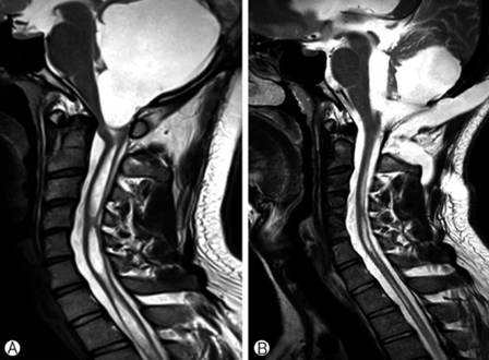
a)Large cystic mass at the posterior fossa with compression of the 4th ventricle b) postoperative MRI, a residual cyst and syringomyelia was decreased.
-
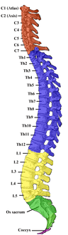
Vertebral column


Other
Surgery is not always recommended for syringomyelia patients. For many patients, the main treatment is analgesia. Physicians specializing in pain management can develop a medication and treatment plan to ameliorate pain. Medications to combat any neuropathic pain symptoms such as shooting and stabbing pains (e.g. gabapentin or pregabalin) would be first-line choices. Opiates are usually prescribed for pain for management of this condition. Facet joint injections are not indicated for the treatment of syringomyelia.[citation needed]
Drugs have no curative value as a treatment for syringomyelia. Radiation is used rarely, and is of little benefit except in the presence of a tumor. In these cases, it can halt the extension of a cavity and may help to alleviate pain.[citation needed]
In the absence of symptoms, syringomyelia is usually not treated. In addition, a physician may recommend not treating the condition in patients of advanced age or in cases where there is no progression of symptoms. Whether treated or not, many patients will be told to avoid activities that involve straining.[citation needed][27]
Since the natural history of syringomyelia is poorly understood, a conservative approach may be recommended. When surgery is not yet advised, patients should be carefully monitored. Periodic MRI's and physical evaluations should be scheduled at the recommendation of a qualified physician.[citation needed]
Research

The precise causes of syringomyelia are still unknown, although blockage of the flow of cerebrospinal fluid has been known to be an important factor since the 1970s. Scientists in the UK and US continue to explore the mechanisms that lead to the formation of syrinxes in the spinal cord. It has been demonstrated that a block of the free flow of cerebrospinal fluid is a contributing factor in the pathogenesis of the disease. Duke University in America and Warwick University are conducting research to explore genetic features of syringomyelia.[28]
Surgical techniques are also being refined by the neurosurgical research community. Successful procedures expand the area around the cerebellum and spinal cord, improving the flow of cerebrospinal fluid and thereby reducing the syrinx.[29]
It is also important to understand the role of birth defects in the development of hindbrain malformations that can lead to syringomyelia, as syringomyelia is a feature of intrauterine life and is also associated with spina bifida. Learning when these defects occur during the development of the fetus can help with the understanding of this and similar disorders, and may lead to preventive treatment that can stop the formation of some birth abnormalities.[citation needed]
Diagnostic technology is another area for continued research. MRI has enabled scientists to see the situation within the spine, including syringomyelia, before any symptoms appear. A new technology, known as dynamic MRI, allows investigators to view spinal fluid flow within the syrinx. CT scans allow physicians to see abnormalities in the brain, and other diagnostic tests have also improved greatly with the availability of new, non-toxic, contrast dyes.[citation needed]
The Chiari & Syringomyelia Foundation, Inc. is a non-profit organization that was founded in October 2007 with the goal of raising awareness and finding a cure for Chiari malformation, syringomyelia and related disorders. In March 2019 plans were announced to work with the family of the famous golfer to re-brand the organization the Bobby Jones Chiari & Syringomyelia Foundation.[citation needed]
See also
- Bobby Jones, a famous golfer, was diagnosed with tumor-related syringomyelia in 1948.
- Brown-Séquard syndrome
- Central cord syndrome
- Dissociated sensory loss
- Ependymoma tumors capable of causing syringomyelia
- Otto Kahler a neurologist in the late 1800s who published the first complete description of syringomyelia.
- Peter McFarline, Australian sports writer who had syringomyelia
- Scoliosis is sometimes caused by syringomyelia.
- Syringobulbia
References
- ↑ "Syringomyelia". Oxford Dictionaries UK Dictionary. Oxford University Press. Retrieved 2016-01-21.
- ↑ "Syringomyelia". Merriam-Webster Dictionary. Retrieved 2016-01-21.
- ↑ Lynn, D. Joanne, Newton, Herbert B. and Rae-Grant, Alexander D. eds. 5-Minute Neurology Consult, The. 2nd Edition. Two Commerce Square, 2001 Market Street, Philadelphia, PA 19103 USA: Lippincott Williams & Wilkins, 2012. Books@Ovid. Web. 03 December, 2020
- ↑ "Neurosurgical considerations in posttraumatic syringomyelia - Home Study Program". AORN Journal. January 2003. Archived from the original on 2012-07-08. Retrieved 2009-02-12.
- ↑ Brewis M, Poskanzer DC, Rolland C, et al., "Neurological disease in an English city". Acta Neurologica Scand Suppl 24:1--89, 1966.
- ↑ Greenberg, David A, et al.: Clinical Neurology. 5th ed. Feb 9, 2002.
- ↑ Nishida, Takayasu, et al. "A large bladder stone caused by syringomyelia" Archived 2007-11-14 at the Wayback Machine. Japanese Journal of Clinical Urology, Vol.60, No. 6, pp 413-415, 2006. ISSN 0385-2393.
- ↑ Hirsch M, et al. Neuropathic osteoarthropathy of the shoulder secondary to syringomyelia. https://doi.org/10.1016/j.diii.2020.09.010 Archived 2022-02-06 at the Wayback Machine
- ↑ "Neuropathic Arthropathy of the Shoulder (Charcot Shoulder): Clinical Commentary" Archived 2020-10-26 at the Wayback Machine Medscape. Accessed 9 January 2011.
- ↑ Byrne. Diseases of the Spine and Spinal Cord.
- ↑ "Arachnoiditis". Archived from the original on 2022-01-25. Retrieved 2022-03-05.
- ↑ https://www.malacards.org/card/syringomyelia#:~:text=the%20initial%20injury.-,Some%20cases%20of%20syringomyelia%20are%20familial%20%2C%20although%20this%20is%20rare,be%20required%20in%20some%20patients Archived 2022-03-02 at the Wayback Machine.
- ↑ Schurch B, Wichmann W, Rossier AB (January 1996). "Post-traumatic syringomyelia (cystic myelopathy): a prospective study of 449 patients with spinal cord injury". J. Neurol. Neurosurg. Psychiatry. 60 (1): 61–7. doi:10.1136/jnnp.60.1.61. PMC 486191. PMID 8558154.
- ↑ "Syringomyelia Fact Sheet | National Institute of Neurological Disorders and Stroke". www.ninds.nih.gov. Archived from the original on 2021-01-19. Retrieved 2021-02-03.
- ↑ Tanghe, H. L. (1995). "Magnetic resonance imaging (MRI) in syringomyelia". Acta Neurochirurgica. 134 (1–2): 93–99. doi:10.1007/BF01428512. hdl:1765/67137. ISSN 0001-6268. PMID 7668137. S2CID 206795042. Archived from the original on 2022-03-19. Retrieved 2022-03-05.
- ↑ "What Is Syringomyelia?". WebMD. Archived from the original on 2021-02-07. Retrieved 2021-02-03.
- ↑ "Syringomyelia". Healthline. 2012-07-23. Archived from the original on 2021-02-07. Retrieved 2021-02-03.
- ↑ Pavaine, Julija; Thompson, Dominic (2019), Barkhof, Frederik; Jäger, Hans Rolf; Thurnher, Majda M.; Rovira, Àlex (eds.), "Imaging of Spinal CSF Disorders: Syringomyelia", Clinical Neuroradiology: The ESNR Textbook, Cham: Springer International Publishing, pp. 519–544, doi:10.1007/978-3-319-68536-6_12, ISBN 978-3-319-68536-6, S2CID 239137707, archived from the original on 2022-03-19, retrieved 2021-02-03
- ↑ "Syringomyelia". NORD (National Organization for Rare Disorders). Archived from the original on 2021-02-05. Retrieved 2021-02-03.
- ↑ "Syringomyelia - an overview | ScienceDirect Topics". www.sciencedirect.com. Archived from the original on 2021-02-10. Retrieved 2021-02-03.
- ↑ "Syringomyelia". Cleveland Clinic. Archived from the original on 2021-02-08. Retrieved 2021-02-03.
- ↑ "Articles". Cedars-Sinai. Archived from the original on 2020-11-20. Retrieved 2021-02-03.
- ↑ "Syringomyelia". www.brainfacts.org. Archived from the original on 2021-02-06. Retrieved 2021-02-03.
- ↑ "Syrinx Symptoms & Treatments | Barrow Neurological Institute". Barrow. Archived from the original on 2022-03-19. Retrieved 2021-02-03.
- ↑ Gottschalk, Andreas; Schmitz, Bernd; Mauer, Uwe M.; Bornstedt, Axel; Steinhoff, Silke; Danz, Burkhardt; Schlötzer, Wiebke; Rasche, Volker (2010). "Dynamic visualization of arachnoid adhesions in a patient with idiopathic syringomyelia using high-resolution cine magnetic resonance imaging at 3T". Journal of Magnetic Resonance Imaging. 32 (1): 218–222. doi:10.1002/jmri.22207. ISSN 1522-2586. PMID 20575079. S2CID 26936003.
- ↑ "Chiari malformation". nhs.uk. 2017-10-18. Archived from the original on 2021-01-25. Retrieved 2021-01-22.
- ↑ National Institute of Neurological Disorders and Stroke. (2021, May 18). Syringomyelia Fact Sheet. https://www.ninds.nih.gov/Disorders/Patient-Caregiver-Education/Fact-Sheets/Syringomyelia-Fact-Sheet
- ↑ ""Information about a Genetic Research Study for Chiari Type I Malformation (CMI) with or without Syringomyelia."". 31 March 2020. Archived from the original on 19 October 2012. Retrieved 5 March 2022.
- ↑ Vandertop, W. (2013-11-22). "Syringomyelia". Neuropediatrics. 45 (1): 003–009. doi:10.1055/s-0033-1361921. ISSN 0174-304X. PMID 24272770. Archived from the original on 2018-06-02. Retrieved 2022-03-19.
External links
| Classification | |
|---|---|
| External resources |
- Pages with script errors
- Webarchive template wayback links
- All articles with unsourced statements
- Articles with unsourced statements from December 2020
- Articles with invalid date parameter in template
- Citation overkill
- Articles tagged with the inline citation overkill template from July 2021
- Articles with unsourced statements from January 2021
- Neurocutaneous conditions
- Rare diseases
- Congenital disorders of nervous system
- Spinal cord disorders