Graves' ophthalmopathy
| Graves ophthalmopathy | |
|---|---|
| Other names: Thyroid eye disease (TED), dysthyroid/thyroid-associated orbitopathy (TAO), Graves' orbitopathy (GO) | |
 | |
| Bulging eyes and lid retraction from Graves' disease | |
Graves ophthalmopathy, also known as thyroid eye disease (TED), is an autoimmune inflammatory disorder of the orbit and periorbital tissues, characterized by upper eyelid retraction, lid lag, swelling, redness (erythema), conjunctivitis, and bulging eyes (exophthalmos).[1] It occurs most commonly in individuals with Graves' disease,[2] and less commonly in individuals with Hashimoto's thyroiditis,[3] or in those who are euthyroid.[4]
It is part of a systemic process with variable expression in the eyes, thyroid, and skin, caused by autoantibodies that bind to tissues in those organs. The autoantibodies target the fibroblasts in the eye muscles, and those fibroblasts can differentiate into fat cells (adipocytes). Fat cells and muscles expand and become inflamed. Veins become compressed, and are unable to drain fluid, causing edema.[1]
Annual incidence is 16/100,000 in women, 3/100,000 in men. About 3–5% have severe disease with intense pain, and sight-threatening corneal ulceration or compression of the optic nerve. Cigarette smoking, which is associated with many autoimmune diseases, raises the incidence 7.7-fold.[1]
Mild disease will often resolve and merely requires measures to reduce discomfort and dryness, such as artificial tears and smoking cessation if possible. Severe cases are a medical emergency, and are treated with glucocorticoids (steroids), and sometimes ciclosporin.[5] Many anti-inflammatory biological mediators, such as infliximab, etanercept, and anakinra are being tried.[1] In January 2020, the US Food and Drug Administration approved teprotumumab-trbw for the treatment of Graves opthalmopathy.[6]
Signs and symptoms
In mild disease, patients present with eyelid retraction. In fact, upper eyelid retraction is the most common ocular sign of Graves' orbitopathy. This finding is associated with lid lag on infraduction (Von Graefe's sign), eye globe lag on supraduction (Kocher's sign), a widened palpebral fissure during fixation (Dalrymple's sign) and an incapacity of closing the eyelids completely (lagophthalmos, Stellwag's sign). Due to the proptosis, eyelid retraction and lagophthalmos, the cornea is more prone to dryness and may present with chemosis, punctate epithelial erosions and superior limbic keratoconjunctivitis. The patients also have a dysfunction of the lacrimal gland with a decrease of the quantity and composition of tears produced. Non-specific symptoms with these pathologies include irritation, grittiness, photophobia, tearing, and blurred vision. Pain is not typical, but patients often complain of pressure in the orbit. Periorbital swelling due to inflammation can also be observed.[citation needed]
- Eye signs[7]
| Sign | Description | Named for |
|---|---|---|
| Abadie's sign | Elevator muscle of upper eyelid is spastic. | Jean Marie Charles Abadie (1842–1932) |
| Ballet's sign | Paralysis of one or more EOM | Louis Gilbert Simeon Ballet (1853–1916) |
| Becker's sign | Abnormal intense pulsation of retina's arteries | Otto Heinrich Enoch Becker (1828–1890) |
| Boston's sign | Jerky movements of upper lid on lower gaze | Leonard Napoleon Boston (1871–1931) |
| Cowen's sign | Extensive hippus of consensual pupillary reflex | Jack Posner Cowen, American ophthalmologist (1906–1989) |
| Dalrymple's sign | Upper eyelid retraction | John Dalrymple (1803–1852) |
| Enroth's sign | Edema esp. of the upper eyelid | Emil Emanuel Enroth, Finnish ophthalmologist (1879–1953) |
| Gifford's sign | Difficulty in eversion of upper lid. | Harold Gifford (1858–1929) |
| Goldzieher's sign | Deep injection of conjunctiva, especially temporal | Wilhelm Goldzieher, Hungarian ophthalmologist (1849–1916) |
| Griffith's sign | Lower lid lag on upward gaze | Alexander James Hill Griffith, English ophthalmologist (1858–1937) |
| Hertoghe's sign | Loss of eyebrows laterally | Eugene Louis Chretien Hertoghe, Dutch thyroid pathologist (1860–1928) |
| Jellinek's sign | Superior eyelid folds is hyperpigmented | Edward Jellinek, English ophthalmologist and pathologist (1890–1963) |
| Joffroy's sign | Absent creases in the fore head on upward gaze. | Alexis Joffroy (1844–1908) |
| Jendrassik's sign | Abduction and rotation of eyeball is limited also | Ernő Jendrassik (1858–1921) |
| Knies's sign | Uneven pupillary dilatation in dim light | Max Knies, German ophthalmologist (1851–1917) |
| Kocher's sign | Spasmatic retraction of upper lid on fixation | Emil Theodor Kocher (1841–1917) |
| Loewi's sign | Quick Mydriasis after instillation of 1:1000 adrenaline | Otto Loewi (1873–1961) |
| Mann's sign | Eyes seem to be situated at different levels because of tanned skin. | John Dixon Mann, English pathologist and forensic scientist (1840–1912) |
| Mean sign | Increased scleral show on upgaze (globe lag) | Named after the expression of being "mean" when viewed from afar, due to the scleral show |
| Möbius's sign | Lack of convergence | Paul Julius Möbius (1853–1907) |
| Payne–Trousseau's sign | Dislocation of globe | John Howard Payne, American surgeon (1916–1983), Armand Trousseau (1801–1867) |
| Pochin's sign | Reduced amplitude of blinking | Sir Edward Eric Pochin (1909–1990) |
| Riesman's sign | Bruit over the eyelid | David Riesman, American physician (1867–1940) |
| Movement's cap phenomenon | Eyeball movements are performed difficultly, abruptly and incompletely | |
| Rosenbach's sign | Eyelids are animated by thin tremors when closed | Ottomar Ernst Felix Rosenbach (1851–1907) |
| Snellen–Riesman's sign | When placing the stethoscope's capsule over closed eyelids a systolic murmur could be heard | Herman Snellen (1834–1908), David Riesman, American physician (1867–1940) |
| Stellwag's sign | Incomplete and infrequent blinking | Karl Stellwag (1823–1904) |
| Suker's sign | Inability to maintain fixation on extreme lateral gaze | George Francis "Franklin" Suker, American ophthalmologist (1869–1933) |
| Topolanski's sign | Around insertion areas of the four rectus muscles of the eyeball a vascular band network is noticed and this network joints the four insertion points. | Alfred Topolanski, Austrian ophthalmologist (1861–1960) |
| von Graefe's sign | Upper lid lag on down gaze | Friedrich Wilhelm Ernst Albrecht von Gräfe (1828–1870) |
| Wilder's sign | Jerking of the eye on movement from abduction to adduction | Helenor Campbell Wilder (née Foerster), American ophthalmologist (1895–1998) |
In moderate active disease, the signs and symptoms are persistent and increasing and include myopathy. The inflammation and edema of the extraocular muscles lead to gaze abnormalities. The inferior rectus muscle is the most commonly affected muscle and patient may experience vertical diplopia on upgaze and limitation of elevation of the eyes due to fibrosis of the muscle. This may also increase the intraocular pressure of the eyes. The double vision is initially intermittent but can gradually become chronic. The medial rectus is the second-most-commonly-affected muscle, but multiple muscles may be affected, in an asymmetric fashion.[citation needed]
In more severe and active disease, mass effects and cicatricial changes occur within the orbit. This is manifested by a progressive exophthalmos, a restrictive myopathy that restricts eye movements and an optic neuropathy. With enlargement of the extraocular muscle at the orbital apex, the optic nerve is at risk of compression. The orbital fat or the stretching of the nerve due to increased orbital volume may also lead to optic nerve damage. The patient experiences a loss of visual acuity, visual field defect, afferent pupillary defect, and loss of color vision. This is an emergency and requires immediate surgery to prevent permanent blindness.[citation needed]
-
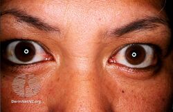
Thyroid ophthalmopathy
-
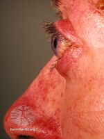
Thyroid ophthalmopathy
-
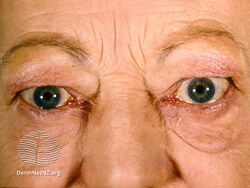
Thyroid ophthalmopathy
-
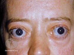
Thyroid ophthalmopathy
.jpg)
.jpg)
.jpg)
.jpg)
Pathophysiology

TAO is an orbital autoimmune disease. The thyroid-stimulating hormone receptor (TSH-R) is an antigen found in orbital fat and connective tissue, and is a target for autoimmune assault.
On histological examination, there is an infiltration of the orbital connective tissue by lymphocytes, plasmocytes, and mastocytes. The inflammation results in a deposition of collagen and glycosaminoglycans in the muscles, which leads to subsequent enlargement and fibrosis. There is also an induction of the lipogenesis by fibroblasts and preadipocytes, which causes enlargement of the orbital fat and extra-ocular muscle compartments. This increase in volume of the intraorbital contents within the confines of the bony orbit may lead to dysthyroid optic neuropathy (DON), increased intraocular pressures, proptosis, and venous congestion leading to chemosis and periorbital oedema.[8][9] In addition, the expansion of the intraorbital soft tissue volume may also remodel the bony orbit and enlarge it, which may be a form of auto-decompression.[10]
Diagnostic
Graves' ophthalmopathy is diagnosed clinically by the presenting ocular signs and symptoms, but positive tests for antibodies (anti-thyroglobulin, anti-microsomal and anti-thyrotropin receptor) and abnormalities in thyroid hormones level (T3, T4, and TSH) help in supporting the diagnosis.[citation needed]
Orbital imaging is an interesting tool for the diagnosis of Graves' ophthalmopathy and is useful in monitoring patients for progression of the disease. It is, however, not warranted when the diagnosis can be established clinically. Ultrasonography may detect early Graves' orbitopathy in patients without clinical orbital findings. It is less reliable than the CT scan and magnetic resonance imaging (MRI), however, to assess the extraocular muscle involvement at the orbital apex, which may lead to blindness. Thus, CT scan or MRI is necessary when optic nerve involvement is suspected. On neuroimaging, the most characteristic findings are thick extraocular muscles with tendon sparing, usually bilateral, and proptosis.[citation needed]
Classification
Mnemonic: "NO SPECS":[11]
| Class | Description |
|---|---|
| Class 0 | No signs or symptoms |
| Class 1 | Only signs (limited to upper lid retraction and stare, with or without lid lag) |
| Class 2 | Soft tissue involvement (oedema of conjunctivae and lids, conjunctival injection, etc.) |
| Class 3 | Proptosis |
| Class 4 | Extraocular muscle involvement (usually with diplopia) |
| Class 5 | Corneal involvement (primarily due to lagophthalmos) |
| Class 6 | Sight loss (due to optic nerve involvement) |
Prevention
Not smoking is a common suggestion in the literature. Apart from smoking cessation, there is little definitive research in this area. In addition to the selenium studies above, some recent research also is suggestive that statin use may assist.[12][13]
Treatment
Even though some people undergo spontaneous remission of symptoms within a year, many need treatment. The first step is the regulation of thyroid hormone levels. Topical lubrication of the eye is used to avoid corneal damage caused by exposure. Corticosteroids are efficient in reducing orbital inflammation, but the benefits cease after discontinuation. Corticosteroids treatment is also limited because of their many side effects. Radiotherapy is an alternative option to reduce acute orbital inflammation. However, there is still controversy surrounding its efficacy. A simple way of reducing inflammation is to stop smoking, as pro-inflammatory substances are found in cigarettes. The medication teprotumumab-trbw may also be used.[14] There is tentative evidence for selenium in mild disease.[15] Tocilizumab, a drug used to suppress the immune system has also been studied as a treatment for TED. However, a Cochrane Review published in 2018 found no evidence (no relevant clinical studies were published) to show that tocilizumab works in people with TED.[16]
In January 2020, the US Food and Drug Administration approved teprotumumab-trbw for the treatment of Graves opthalmopathy.[6]
Surgery
There is some evidence that a total or sub-total thyroidectomy may assist in reducing levels of TSH receptor antibodies (TRAbs) and as a consequence reduce the eye symptoms, perhaps after a 12-month lag.[17][12][18][19][20] However, a 2015 meta review found no such benefits,[21] and there is some evidence that suggests that surgery is no better than medication.[22]
Surgery may be done to decompress the orbit, to improve the proptosis, and to address the strabismus causing diplopia. Surgery is performed once the person's disease has been stable for at least six months. In severe cases, however, the surgery becomes urgent to prevent blindness from optic nerve compression. Because the eye socket is bone, there is nowhere for eye muscle swelling to be accommodated, and, as a result, the eye is pushed forward into a protruded position. Orbital decompression involves removing some bone from the eye socket to open up one or more sinuses and so make space for the swollen tissue and allowing the eye to move back into normal position and also relieving compression of the optic nerve that can threaten sight.
Eyelid surgery is the most common surgery performed on Graves ophthalmopathy patients. Lid-lengthening surgeries can be done on upper and lower eyelid to correct the patient's appearance and the ocular surface exposure symptoms. Marginal myotomy of levator palpebrae muscle can reduce the palpebral fissure height by 2–3 mm. When there is a more severe upper lid retraction or exposure keratitis, marginal myotomy of levator palpebrae associated with lateral tarsal canthoplasty is recommended. This procedure can lower the upper eyelid by as much as 8 mm. Other approaches include müllerectomy (resection of the Müller muscle), eyelid spacer grafts, and recession of the lower eyelid retractors. Blepharoplasty can also be done to debulk the excess fat in the lower eyelid.[23]
A summary of treatment recommendations was published in 2015 by an Italian taskforce,[24] which largely supports the other studies.
Prognosis
Risk factors of progressive and severe thyroid-associated orbitopathy are:[citation needed]
- Age greater than 50 years
- Rapid onset of symptoms under 3 months
- Cigarette smoking
- Diabetes
- Severe or uncontrolled hyperthyroidism
- Presence of pretibial myxedema
- High cholesterol levels (hyperlipidemia)
- Peripheral vascular disease
Epidemiology
The pathology mostly affects persons of 30 to 50 years of age. Females are four times more likely to develop TAO than males. When males are affected, they tend to have a later onset and a poor prognosis. A study demonstrated that at the time of diagnosis, 90% of the patients with clinical orbitopathy were hyperthyroid according to thyroid function tests, while 3% had Hashimoto's thyroiditis, 1% were hypothyroid and 6% did not have any thyroid function tests abnormality.[25] Of patients with Graves' hyperthyroidism, 20 to 25 percent have clinically obvious Graves' ophthalmopathy, while only 3–5% will develop severe ophthalmopathy.[26][27]
History
In medical literature, Robert James Graves, in 1835, was the first to describe the association of a thyroid goitre with exophthalmos (proptosis) of the eye.[28] Graves' ophthalmopathy may occur before, with, or after the onset of overt thyroid disease and usually has a slow onset over many months.
See also
References
- ↑ 1.0 1.1 1.2 1.3 Bahn, Rebecca S. (2010). "Graves' Ophthalmopathy". New England Journal of Medicine. 362 (8): 726–38. doi:10.1056/NEJMra0905750. PMC 3902010. PMID 20181974.
- ↑ Wiersinga, Wilmar M.; Bartalena, Luigi (October 2002). "Epidemiology and prevention of Graves' ophthalmopathy". Thyroid. 12 (10): 855–860. doi:10.1089/105072502761016476. ISSN 1050-7256. PMID 12487767.
- ↑ Kan, Emrah; Kan, Elif Kilic; Ecemis, Gülcin; Colak, Ramis (2014-08-18). "Presence of thyroid-associated ophthalmopathy in Hashimoto's thyroiditis". International Journal of Ophthalmology. 7 (4): 644–647. doi:10.3980/j.issn.2222-3959.2014.04.10. ISSN 2222-3959. PMC 4137199. PMID 25161935.
- ↑ Solomon, David H.; Chopra, Inder J.; Chopra, Usha; Smith, Francoise J. (1977-01-27). "Identification of Subgroups of Euthyroid Graves's Ophthalmopathy". New England Journal of Medicine. 296 (4): 181–186. doi:10.1056/nejm197701272960401. ISSN 0028-4793. PMID 576175.
- ↑ Harrison's Principles of Internal Medicine, 16th Ed., Ch. 320, Disorders of the Thyroid Gland
- ↑ 6.0 6.1 Commissioner, Office of the (2020-03-24). "FDA approves first treatment for thyroid eye disease". FDA. Archived from the original on 2021-11-26. Retrieved 2021-02-06.
- ↑ "Archive copy". Archived from the original on 2014-04-17. Retrieved 2013-09-04.
{{cite web}}: CS1 maint: archived copy as title (link)[full citation needed] - ↑ Feldon, S. E.; Muramatsu, S.; Weiner, J. M. (October 1984). "Clinical classification of Graves' ophthalmopathy. Identification of risk factors for optic neuropathy". Archives of Ophthalmology. 102 (10): 1469–1472. doi:10.1001/archopht.1984.01040031189015. ISSN 0003-9950. PMID 6548373.
- ↑ Ohtsuka, K. (October 1997). "Intraocular pressure and proptosis in 95 patients with Graves ophthalmopathy". American Journal of Ophthalmology. 124 (4): 570–572. doi:10.1016/s0002-9394(14)70883-9. ISSN 0002-9394. PMID 9323958.
- ↑ Tan, Nicholas Y. Q.; Leong, Yuan-Yuh; Lang, Stephanie S.; Htoon, Zin M.; Young, Stephanie M.; Sundar, Gangadhara (2017-05-01). "Radiologic Parameters of Orbital Bone Remodeling in Thyroid Eye DiseaseOrbital Bone Remodeling in Thyroid Eye Disease". Investigative Ophthalmology & Visual Science. 58 (5): 2527–2533. doi:10.1167/iovs.16-21035. ISSN 1552-5783. PMID 28492870.
- ↑ Cawood, T.; Moriarty, P; O'Shea, D (2004). "Recent developments in thyroid eye disease". BMJ. 329 (7462): 385–90. doi:10.1136/bmj.329.7462.385. PMC 509348. PMID 15310608.
- ↑ 12.0 12.1 De Bellis, Annamaria; Conzo, Giovanni; Cennamo, Gilda; Pane, Elena; Bellastella, Giuseppe; Colella, Caterina; Iacovo, Assunta Dello; Paglionico, Vanda Amoresano; Sinisi, Antonio Agostino; Wall, Jack R.; Bizzarro, Antonio; Bellastella, Antonio (2011). "Time course of Graves' ophthalmopathy after total thyroidectomy alone or followed by radioiodine therapy: A 2-year longitudinal study". Endocrine. 41 (2): 320–6. doi:10.1007/s12020-011-9559-x. PMID 22169963. S2CID 8197441.
- ↑ Kuehn, Bridget M. (December 15, 2014). "Surgery, Statins Linked to Lower Graves' Complication Risk". Medscape Medical News. Archived from the original on December 28, 2014. Retrieved January 15, 2015.
- ↑ "FDA approves first treatment for thyroid eye disease". FDA. 21 January 2020. Archived from the original on 26 November 2021. Retrieved 27 January 2020.
- ↑ Ruchała, M; Sawicka-Gutaj, N (July 2016). "Advances in the pharmacological treatment of Graves' orbitopathy". Expert Review of Clinical Pharmacology. 9 (7): 981–9. doi:10.1586/17512433.2016.1165606. PMID 26966785. S2CID 9780703.
- ↑ Hamed Azzam, Shirin; Kang, Swan; Salvi, Mario; Ezra, Daniel G (2018-11-27). Cochrane Eyes and Vision Group (ed.). "Tocilizumab for thyroid eye disease". Cochrane Database of Systematic Reviews. 11: CD012984. doi:10.1002/14651858.CD012984.pub2. PMC 6517231. PMID 30480323.
- ↑ Takamura, Yuuki; Nakano, Keiichi; Uruno, Takashi; Ito, Yasuhiro; Miya, Akihiro; Kobayashi, Kaoru; Yokozawa, Tamotsu; Matsuzuka, Fumio; Kuma, Kanji; Miyauchi, Akira (2003). "Changes in Serum TSH Receptor Antibody (TRAb) Values in Patients with Graves' Disease after Total or Subtotal Thyroidectomy". Endocrine Journal. 50 (5): 595–601. doi:10.1507/endocrj.50.595. PMID 14614216.
- ↑ Bhargav, P. R. K.; Sabaretnam, M.; Kumar, S. Chandra; Zwalitha, S.; Devi, N. Vimala (2016-06-22). "Regression of Ophthalmopathic Exophthalmos in Graves' Disease After Total Thyroidectomy: a Prospective Study of a Surgical Series". Indian Journal of Surgery. 79 (6): 521–526. doi:10.1007/s12262-016-1516-8. ISSN 0972-2068. PMC 5711711. PMID 29217903.
- ↑ Nart, Ahmet; Uslu, Adam; Aykas, Ahmet; Yüzbaşoğlu, Fatih; Doğan, Murat; Demirtaş, Özgür; Şimşek, Cenk (2008-07-01). "Total Thyroidectomy for the Treatment of Recurrent Graves' Disease With Ophthalmopathy". Asian Journal of Surgery. 31 (3): 115–118. doi:10.1016/S1015-9584(08)60070-6. PMID 18658008.
- ↑ Lowery, A. J.; Kerin, M. J. (2009-10-01). "Graves' ophthalmopathy: the case for thyroid surgery". The Surgeon. 7 (5): 290–296. doi:10.1016/s1479-666x(09)80007-3. ISSN 1479-666X. PMID 19848063.
- ↑ Liu, Zi Wei; Masterson, Liam; Fish, Brian; Jani, Piyush; Chatterjee, Krishna (2015-11-25). "Thyroid surgery for Graves' disease and Graves' ophthalmopathy". The Cochrane Database of Systematic Reviews (11): CD010576. doi:10.1002/14651858.CD010576.pub2. ISSN 1469-493X. PMID 26606533.
- ↑ Laurberg, P.; Wallin, G.; Tallstedt, L.; Abraham-Nordling, M.; Lundell, G.; Torring, O. (2007). "TSH-receptor autoimmunity in Graves' disease after therapy with anti-thyroid drugs, surgery, or radioiodine: A 5-year prospective randomized study". European Journal of Endocrinology. 158 (1): 69–75. doi:10.1530/EJE-07-0450. PMID 18166819.
- ↑ Muratet JM. "Eyelid retraction". Ophthalmic Plastic Surgery. Le Syndicat National des Ophtalmologistes de France. Archived from the original on June 9, 2007. Retrieved 2007-07-12.
- ↑ Bartalena, L.; MacChia, P. E.; Marcocci, C.; Salvi, M.; Vermiglio, F. (2015). "Effects of treatment modalities for Graves' hyperthyroidism on Graves' orbitopathy: A 2015 Italian Society of Endocrinology Consensus Statement". Journal of Endocrinological Investigation. 38 (4): 481–7. doi:10.1007/s40618-015-0257-z. PMC 4374116. PMID 25722226.
- ↑ Bartley, G. B.; Fatourechi, V; Kadrmas, E. F.; Jacobsen, S. J.; Ilstrup, D. M.; Garrity, J. A.; Gorman, C. A. (1996). "Clinical features of Graves' ophthalmopathy in an incidence cohort". American Journal of Ophthalmology. 121 (3): 284–90. doi:10.1016/s0002-9394(14)70276-4. PMID 8597271.
- ↑ Davies, Terry F (September 2009). Ross, Douglas S; Martin, Kathryn A (eds.). "Pathogenesis and clinical features of Graves' ophthalmopathy (orbitopathy)". UpToDate. Archived from the original on 2015-05-18. Retrieved 2015-05-08.
- ↑ Bartalena, L; Marcocci, C; Pinchera, A (2002). "Graves' ophthalmopathy: A preventable disease?". European Journal of Endocrinology. 146 (4): 457–61. doi:10.1530/eje.0.1460457. PMID 11916611.
- ↑ Robert James Graves at Who Named It?
Further reading
- Behbehani, Raed; Sergott, Robert C; Savino, Peter J (2004). "Orbital radiotherapy for thyroid-related orbitopathy". Current Opinion in Ophthalmology. 15 (6): 479–82. doi:10.1097/01.icu.0000144388.89867.03. PMID 15523191. S2CID 31340321.
- Boncoeur, M.-P. (2004). "Orbitopathie dysthyroïdienne : imagerie : Orbitopathie dysthyroïdienne" [Imaging techniques in Graves disease : Dysthyroid orbitopathy]. Journal Français d'Ophtalmologie (in français). 27 (7): 815–8. doi:10.1016/S0181-5512(04)96221-3. PMID 15499283. INIST:16100159.
- Boulos, Patrick Roland; Hardy, Isabelle (2004). "Thyroid-associated orbitopathy: A clinicopathologic and therapeutic review". Current Opinion in Ophthalmology. 15 (5): 389–400. doi:10.1097/01.icu.0000139992.15463.1b. PMID 15625899. S2CID 23194226.
- Camezind, P.; Robert, P.-Y.; Adenis, J.-P. (2004). "Signes cliniques de l'orbitopathie dysthyroïdienne : Orbitopathie dysthyroïdienne" [Clinical signs of dysthyroid orbitopathy : Dysthyroid orbitopathy]. Journal Français d'Ophtalmologie (in français). 27 (7): 810–4. doi:10.1016/S0181-5512(04)96220-1. PMID 15499282. INIST:16100158.
- Duker, Jay S.; Yanoff, Myron (2004). "chapt 95". Ophthalmology (2nd ed.). Saint Louis: C.V. Mosby. ISBN 978-0-323-02907-0.
- Morax, S.; Ben Ayed, H. (2004). "Techniques et indications chirurgicales des décompressions osseuses de l'orbitopathie dysthyroïdienne" [Orbital decompression for dysthyroid orbitopathy: a review of techniques and indications]. Journal Français d'Ophtalmologie (in français). 27 (7): 828–44. doi:10.1016/s0181-5512(04)96225-0. PMID 15499287.
- Rose, John G.; Burkat, Cat Nguyen; Boxrud, Cynthia A. (2005). "Diagnosis and Management of Thyroid Orbitopathy". Otolaryngologic Clinics of North America. 38 (5): 1043–74. doi:10.1016/j.otc.2005.03.015. PMID 16214573.
External links
| Classification | |
|---|---|
| External resources |
- Pages with script errors
- CS1 maint: archived copy as title
- All articles with incomplete citations
- Articles with incomplete citations from May 2015
- Articles with invalid date parameter in template
- All articles with unsourced statements
- Articles with unsourced statements from August 2020
- CS1 français-language sources (fr)
- Thyroid disease
- Diseases of the eye and adnexa
- Autoimmune diseases
- Thyroid