GRIK1
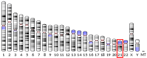
Glutamate receptor, ionotropic, kainate 1, also known as GRIK1, is a protein that in humans is encoded by the GRIK1 gene.[5]
Function
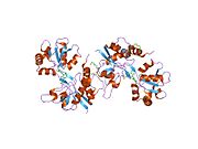
This gene encodes one of the many ionotropic glutamate receptor (GluR) subunits that function as a ligand-gated ion channel. The specific GluR subunit encoded by this gene is of the kainate receptor subtype. Receptor assembly and intracellular trafficking of ionotropic glutamate receptors are regulated by RNA editing and alternative splicing. These receptors mediate excitatory neurotransmission and are critical for normal synaptic function. Two alternatively spliced transcript variants that encode different isoforms have been described. Exons of this gene are interspersed with exons from the C21orf41 gene, which is transcribed in the same orientation as this gene but does not seem to encode a protein.[5]
Interactions
GRIK1 has been shown to interact with DLG4,[6] PICK1[6] and SDCBP.[6]
RNA editing
Type
A to I RNA editing is catalyzed by a family of adenosine deaminases acting on RNA (ADARs) that specifically recognize adenosines within double-stranded regions of pre-mRNAs and deaminate them to inosine. Inosines are recognised as guanosine by the cells translational machinery. There are three members of the ADAR family ADARs 1-3, with ADAR1 and ADAR2 being the only enzymatically active members. ADAR3 is thought to have a regulatory role in the brain. ADAR1 and ADAR2 are widely expressed in tissues, whereas ADAR3 is restricted to the brain. The double-stranded regions of RNA are formed by base-pairing between residues in the close to region of the editing site, with residues usually in a neighboring intron, but can be an exonic sequence. The region that base-pairs with the editing region is known as an Editing Complementary Sequence (ECS). ADARs bind interact directly with the dsRNA substrate via their double-stranded RNA binding domains. If an editing site occurs within a coding sequence, the result could be a codon change. This can lead to translation of a protein isoform due to a change in its primary protein structure. Therefore, editing can also alter protein function. A to I editing occurs in a noncoding RNA sequences such as introns, untranslated regions (UTRs), LINEs, SINEs( especially Alu repeats). The function of A to I editing in these regions is thought to involve creation of splice sites and retention of RNAs in the nucleus, among others.
Location
The pre-mRNA of GluR-5 is edited at one position at the Q/R site located at membrane region 2 (M2). There is a codon change as a result of editing. The codon change is (CAG) Glutamine (Q) to (CGG) an Arginine (R).[7] Like GluR-6 the ECS is located about 2000 nucleotides downstream of the editing site.[8]
Regulation
Editing of the Q/R site is development- and tissue-regulated. Editing in the spinal cord, corpus callosum, cerebellum is 50%, while editing in the Thalamus, amygdala, hippocampus is about 70%.
Consequences
Structure
Editing results in a change in amino acid in the second membrane domain of the receptor.
Function
The editing site is found within the second intracellular domain. It is thought that editing affects the permeability of the receptor to CA2+. Editing of the Q/R site is thought to reduce the permeability of the channel to Ca2+[7]
RNA editing of the Q/R site can effect inhibition of the channel by membrane fatty acids such as arachidonic acid and docosahexaenoic acid[9] For Kainate receptors with only edited isoforms, these are strongly inhibited by these fatty acids. However, inclusion of just one nonedited subunit is enough to stop this inhibition(.[9]
See also
- Kainate receptor
- GRIK1 RNA editing
References
- ^ a b c GRCh38: Ensembl release 89: ENSG00000171189 – Ensembl, May 2017
- ^ a b c GRCm38: Ensembl release 89: ENSMUSG00000022935 – Ensembl, May 2017
- ^ "Human PubMed Reference:". National Center for Biotechnology Information, U.S. National Library of Medicine.
- ^ "Mouse PubMed Reference:". National Center for Biotechnology Information, U.S. National Library of Medicine.
- ^ a b "Entrez Gene: GRIK1 glutamate receptor, ionotropic, kainate 1".
- ^ a b c Hirbec, Hélène; Francis, Joanna C.; Lauri, Sari E.; Braithwaite, Steven P.; Coussen, Françoise; Mulle, Christophe; Dev, Kumlesh K.; Coutinho, Victoria; Meyer, Guido; Isaac, John T. R.; Collingridge, Graham L.; Henley, Jeremy M.; Couthino, Victoria (Feb 2003). "Rapid and differential regulation of AMPA and kainate receptors at hippocampal mossy fibre synapses by PICK1 and GRIP". Neuron. 37 (4). United States: 625–38. doi:10.1016/S0896-6273(02)01191-1. ISSN 0896-6273. PMC 3314502. PMID 12597860.
- ^ a b Seeburg PH, Single F, Kuner T, Higuchi M, Sprengel R (July 2001). "Genetic manipulation of key determinants of ion flow in glutamate receptor channels in the mouse". Brain Res. 907 (1–2): 233–43. doi:10.1016/S0006-8993(01)02445-3. PMID 11430906. S2CID 11969068.
- ^ Herb A, Higuchi M, Sprengel R, Seeburg PH (March 1996). "Q/R site editing in kainate receptor GluR5 and GluR6 pre-mRNAs requires distant intronic sequences". Proc. Natl. Acad. Sci. U.S.A. 93 (5): 1875–80. Bibcode:1996PNAS...93.1875H. doi:10.1073/pnas.93.5.1875. PMC 39875. PMID 8700852.
- ^ a b Wilding TJ, Fulling E, Zhou Y, Huettner JE (July 2008). "Amino Acid Substitutions in the Pore Helix of GluR6 Control Inhibition by Membrane Fatty Acids". J. Gen. Physiol. 132 (1): 85–99. doi:10.1085/jgp.200810009. PMC 2442176. PMID 18562501.
Further reading
- Nutt SL, Kamboj RK (1995). "RNA editing of human kainate receptor subunits". NeuroReport. 5 (18): 2625–9. doi:10.1097/00001756-199412000-00055. PMID 7696618.
- Roche KW, Raymond LA, Blackstone C, Huganir RL (1994). "Transmembrane topology of the glutamate receptor subunit GluR6". J. Biol. Chem. 269 (16): 11679–82. doi:10.1016/S0021-9258(17)32623-6. PMID 8163463.
- Gregor P, O'Hara BF, Yang X, Uhl GR (1994). "Expression and novel subunit isoforms of glutamate receptor genes GluR5 and GluR6". NeuroReport. 4 (12): 1343–6. doi:10.1097/00001756-199309150-00014. PMID 8260617.
- Eubanks JH, Puranam RS, Kleckner NW, et al. (1993). "The gene encoding the glutamate receptor subunit GluR5 is located on human chromosome 21q21.1-22.1 in the vicinity of the gene for familial amyotrophic lateral sclerosis". Proc. Natl. Acad. Sci. U.S.A. 90 (1): 178–82. Bibcode:1993PNAS...90..178E. doi:10.1073/pnas.90.1.178. PMC 45623. PMID 8419920.
- Potier MC, Dutriaux A, Lambolez B, et al. (1993). "Assignment of the human glutamate receptor gene GLUR5 to 21q22 by screening a chromosome 21 YAC library". Genomics. 15 (3): 696–7. doi:10.1006/geno.1993.1131. PMID 8468067.
- Korczak B, Nutt SL, Fletcher EJ, et al. (1996). "cDNA cloning and functional properties of human glutamate receptor EAA3 (GluR5) in homomeric and heteromeric configuration". Recept. Channels. 3 (1): 41–9. PMID 8589992.
- Brakeman PR, Lanahan AA, O'Brien R, et al. (1997). "Homer: a protein that selectively binds metabotropic glutamate receptors". Nature. 386 (6622): 284–8. Bibcode:1997Natur.386..284B. doi:10.1038/386284a0. PMID 9069287. S2CID 4346579.
- Sander T, Hildmann T, Kretz R, et al. (1997). "Allelic association of juvenile absence epilepsy with a GluR5 kainate receptor gene (GRIK1) polymorphism". Am. J. Med. Genet. 74 (4): 416–21. doi:10.1002/(SICI)1096-8628(19970725)74:4<416::AID-AJMG13>3.0.CO;2-L. PMID 9259378.
- Clarke VR, Ballyk BA, Hoo KH, et al. (1997). "A hippocampal GluR5 kainate receptor regulating inhibitory synaptic transmission". Nature. 389 (6651): 599–603. Bibcode:1997Natur.389..599C. doi:10.1038/39315. PMID 9335499. S2CID 4426839.
- Xiao B, Tu JC, Petralia RS, et al. (1998). "Homer regulates the association of group 1 metabotropic glutamate receptors with multivalent complexes of homer-related, synaptic proteins". Neuron. 21 (4): 707–16. doi:10.1016/S0896-6273(00)80588-7. PMID 9808458. S2CID 16431031.
- Montague AA, Greer CA (1999). "Differential distribution of ionotropic glutamate receptor subunits in the rat olfactory bulb". J. Comp. Neurol. 405 (2): 233–46. doi:10.1002/(SICI)1096-9861(19990308)405:2<233::AID-CNE7>3.0.CO;2-A. PMID 10023812. S2CID 1317987.
- Bortolotto ZA, Clarke VR, Delany CM, et al. (1999). "Kainate receptors are involved in synaptic plasticity". Nature. 402 (6759): 297–301. Bibcode:1999Natur.402..297B. doi:10.1038/46290. PMID 10580501. S2CID 205051834.
- Barbon A, Barlati S (2000). "Genomic organization, proposed alternative splicing mechanisms, and RNA editing structure of GRIK1". Cytogenet. Cell Genet. 88 (3–4): 236–9. doi:10.1159/000015558. PMID 10828597. S2CID 5850944.
- Hattori M, Fujiyama A, Taylor TD, et al. (2000). "The DNA sequence of human chromosome 21". Nature. 405 (6784): 311–9. Bibcode:2000Natur.405..311H. doi:10.1038/35012518. PMID 10830953.
- Ango F, Prézeau L, Muller T, et al. (2001). "Agonist-independent activation of metabotropic glutamate receptors by the intracellular protein Homer". Nature. 411 (6840): 962–5. Bibcode:2001Natur.411..962A. doi:10.1038/35082096. PMID 11418862. S2CID 4417727.
- Shibata H, Joo A, Fujii Y, et al. (2002). "Association study of polymorphisms in the GluR5 kainate receptor gene (GRIK1) with schizophrenia". Psychiatr. Genet. 11 (3): 139–44. doi:10.1097/00041444-200109000-00005. PMID 11702055. S2CID 38356907.
- Hirbec H, Perestenko O, Nishimune A, et al. (2002). "The PDZ proteins PICK1, GRIP, and syntenin bind multiple glutamate receptor subtypes. Analysis of PDZ binding motifs" (PDF). J. Biol. Chem. 277 (18): 15221–4. doi:10.1074/jbc.C200112200. PMID 11891216. S2CID 13732968.
- Enz R (2002). "The actin-binding protein Filamin-A interacts with the metabotropic glutamate receptor type 7". FEBS Lett. 514 (2–3): 184–8. doi:10.1016/S0014-5793(02)02361-X. PMID 11943148. S2CID 44474808.
- Hirbec H, Francis JC, Lauri SE, et al. (2003). "Rapid and differential regulation of AMPA and kainate receptors at hippocampal mossy fibre synapses by PICK1 and GRIP". Neuron. 37 (4): 625–38. doi:10.1016/S0896-6273(02)01191-1. PMC 3314502. PMID 12597860.
- Ren Z, Riley NJ, Needleman LA, et al. (2004). "Cell surface expression of GluR5 kainate receptors is regulated by an endoplasmic reticulum retention signal". J. Biol. Chem. 278 (52): 52700–9. doi:10.1074/jbc.M309585200. PMID 14527949.
External links
- GRIK1+protein,+human at the U.S. National Library of Medicine Medical Subject Headings (MeSH)
This article incorporates text from the United States National Library of Medicine, which is in the public domain.


