File:Pulmonary-cryptococcosis-6.png
Jump to navigation
Jump to search

Size of this preview: 578 × 600 pixels. Other resolutions: 231 × 240 pixels | 463 × 480 pixels | 740 × 768 pixels | 987 × 1,024 pixels | 1,974 × 2,048 pixels | 2,762 × 2,866 pixels.
Original file (2,762 × 2,866 pixels, file size: 2.83 MB, MIME type: image/png)
Summary
Author: Case courtesy of Dr Henry Knipe, Radiopaedia.org, rID: 53233 Source:https://radiopaedia.org/cases/pulmonary-cryptococcosis-6?lang=gb
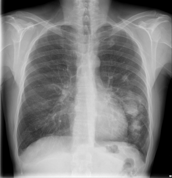
Description:X-ray chest showing Nodular densities project over the left mid to lower zones. Right lung is clear. Cardiomediastinal contour is normal. Biopsy of lingular of lung: Cryptococcosis
Licensing
| This work is licensed under the Creative Commons Attribution-NonCommersial-ShareAlike 4.0 License. |
File history
Click on a date/time to view the file as it appeared at that time.
| Date/Time | Thumbnail | Dimensions | User | Comment | |
|---|---|---|---|---|---|
| current | 07:02, 7 June 2021 | | 2,762 × 2,866 (2.83 MB) | Whispyhistory (talk | contribs) | Author: Case courtesy of Dr Henry Knipe, Radiopaedia.org, rID: 53233 Source:https://radiopaedia.org/cases/pulmonary-cryptococcosis-6?lang=gb Description:X-ray chest showing Nodular densities project over the left mid to lower zones. Right lung is clear. Cardiomediastinal contour is normal. Biopsy of lingular of lung: Cryptococcosis |
You cannot overwrite this file.
File usage
The following file is a duplicate of this file (more details):
- File:Pulmonary cryptococcosis (Radiopaedia 53233-59203 Frontal 1).png from a shared repository
.png)
The following 2 pages use this file: