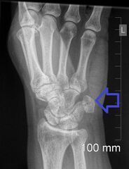
File:Pisiform fracture (Radiopaedia 52465).jpeg
Jump to navigation
Jump to search

Size of this preview: 459 × 600 pixels. Other resolutions: 184 × 240 pixels | 466 × 609 pixels.
Original file (466 × 609 pixels, file size: 87 KB, MIME type: image/jpeg)
.jpeg)
File history
Click on a date/time to view the file as it appeared at that time.
| Date/Time | Thumbnail | Dimensions | User | Comment | |
|---|---|---|---|---|---|
| current | 06:09, 24 September 2022 | | 466 × 609 (87 KB) | Doc James | Added arrow |
| 12:27, 26 March 2021 | 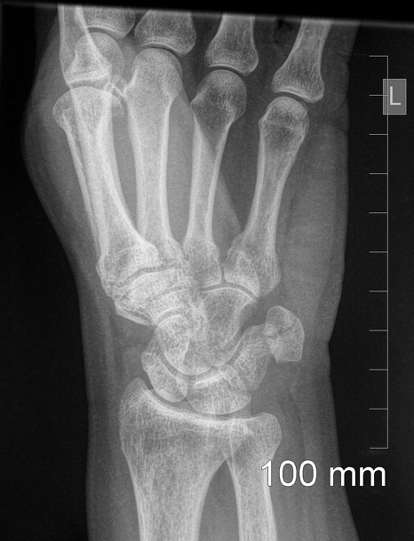 | 466 × 609 (171 KB) | Fæ | Radiopaedia project rID:52465 (batch #29414) |
File usage
There are no pages that use this file.
.jpeg)