File:Periosteal-osteosarcoma-1.jpg
Jump to navigation
Jump to search

Size of this preview: 240 × 599 pixels. Other resolutions: 96 × 240 pixels | 192 × 480 pixels | 836 × 2,088 pixels.
Original file (836 × 2,088 pixels, file size: 382 KB, MIME type: image/jpeg)
Summary
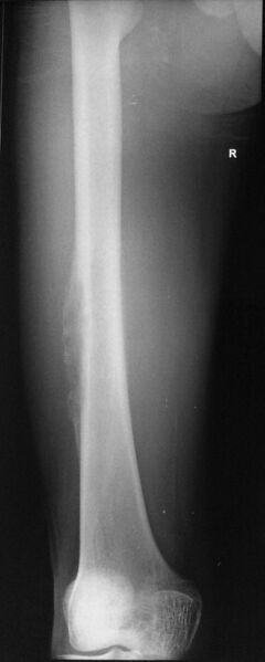
Author:Case courtesy of Dr Subash Thapa, Radiopaedia.org, rID: 40937 Source:https://radiopaedia.org/cases/periosteal-osteosarcoma-1?lang=gb Description:X-ray femur. There is a broad based eccentric lytic lesion in the diaphysis of the right midshaft of femur involving the lateral cortex with periosteal reaction. There is also evidence of thickening of the adjacent cortex. Soft tissue opacity mass is seen anteriorly, suggesting an extraosseous soft tissue component.
Licensing
| This work is licensed under the Creative Commons Attribution-NonCommersial-ShareAlike 4.0 License. |
File history
Click on a date/time to view the file as it appeared at that time.
| Date/Time | Thumbnail | Dimensions | User | Comment | |
|---|---|---|---|---|---|
| current | 10:33, 5 July 2021 | 836 × 2,088 (382 KB) | Whispyhistory (talk | contribs) | Author:Case courtesy of Dr Subash Thapa, Radiopaedia.org, rID: 40937 Source:https://radiopaedia.org/cases/periosteal-osteosarcoma-1?lang=gb Description:X-ray femur. There is a broad based eccentric lytic lesion in the diaphysis of the right midshaft of femur involving the lateral cortex with periosteal reaction. There is also evidence of thickening of the adjacent cortex. Soft tissue opacity mass is seen anteriorly, suggesting an extraosseous soft tissue component. |
You cannot overwrite this file.
File usage
The following file is a duplicate of this file (more details):
- File:Periosteal osteosarcoma (Radiopaedia 40937-43646 Frontal 1).jpg from a shared repository
There are no pages that use this file.