File:Periosteal-chondroma-1.png

Original file (836 × 868 pixels, file size: 468 KB, MIME type: image/png)
Summary
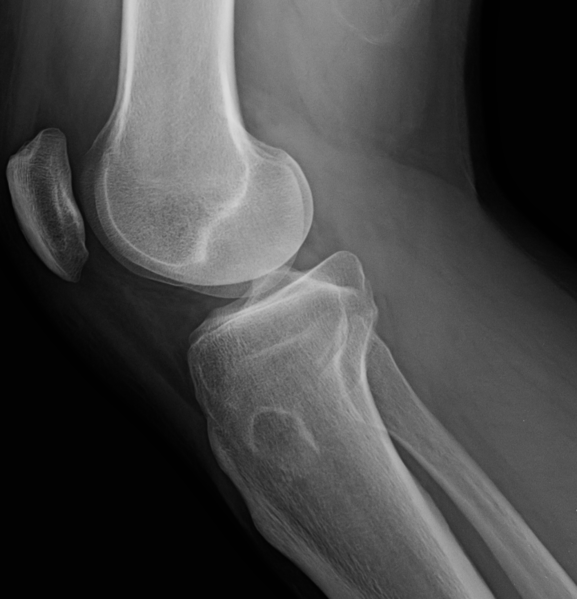
Author: Case courtesy of Dr Brian Gilcrease-Garcia, Radiopaedia.org, rID: 56933 Source:https://radiopaedia.org/cases/periosteal-chondroma-1?lang=gb Description: X-ray knee (lateral view)- Right knee radiographs (age-25) show an eccentric, cortically-based lytic lesion located at the anterolateral right proximal tibia (best visualised on oblique and lateral views). There is a small amount of internal chondroid calcification. The joint spaces of the knee are normal. No knee effusion, and no acute fracture. The small lytic mass associated with the anterolateral cortex of the tibia metaphysis shows evidence of chondroid-type mineralisation by radiography ("rings and arcs" pattern of calcification). MRI images nicely demonstrate the lesion originating from the superficial cortex, without intramedullary invasion. The absence of associated reactive marrow oedema is suggestive of a benign aetiology. The MR signal characteristics (T1 intermediate, T2 hyperintense) are typical of chondral tissue.
Licensing
| This work is licensed under the Creative Commons Attribution-NonCommersial-ShareAlike 4.0 License. |
File history
Click on a date/time to view the file as it appeared at that time.
| Date/Time | Thumbnail | Dimensions | User | Comment | |
|---|---|---|---|---|---|
| current | 05:19, 15 June 2021 | | 836 × 868 (468 KB) | Whispyhistory (talk | contribs) | Author: Case courtesy of Dr Brian Gilcrease-Garcia, Radiopaedia.org, rID: 56933 Source:https://radiopaedia.org/cases/periosteal-chondroma-1?lang=gb Description: X-ray knee (lateral view)- Right knee radiographs (age-25) show an eccentric, cortically-based lytic lesion located at the anterolateral right proximal tibia (best visualised on oblique and lateral views). There is a small amount of internal chondroid calcification. The joint spaces of the knee are normal. No knee effusion, and no acu... |
You cannot overwrite this file.
File usage
The following file is a duplicate of this file (more details):
- File:Periosteal chondroma (Radiopaedia 56933-63776 Lateral 1).png from a shared repository
.png)
The following 2 pages use this file: