File:Parasite140076-fig1 Dirofilaria repens removed from a subcutaneous nodule - Photos.png
Jump to navigation
Jump to search
Size of this preview: 341 × 600 pixels. Other resolutions: 136 × 240 pixels | 273 × 480 pixels | 436 × 768 pixels | 582 × 1,024 pixels | 1,645 × 2,894 pixels.
Original file (1,645 × 2,894 pixels, file size: 5.31 MB, MIME type: image/png)
Summary
| Description |
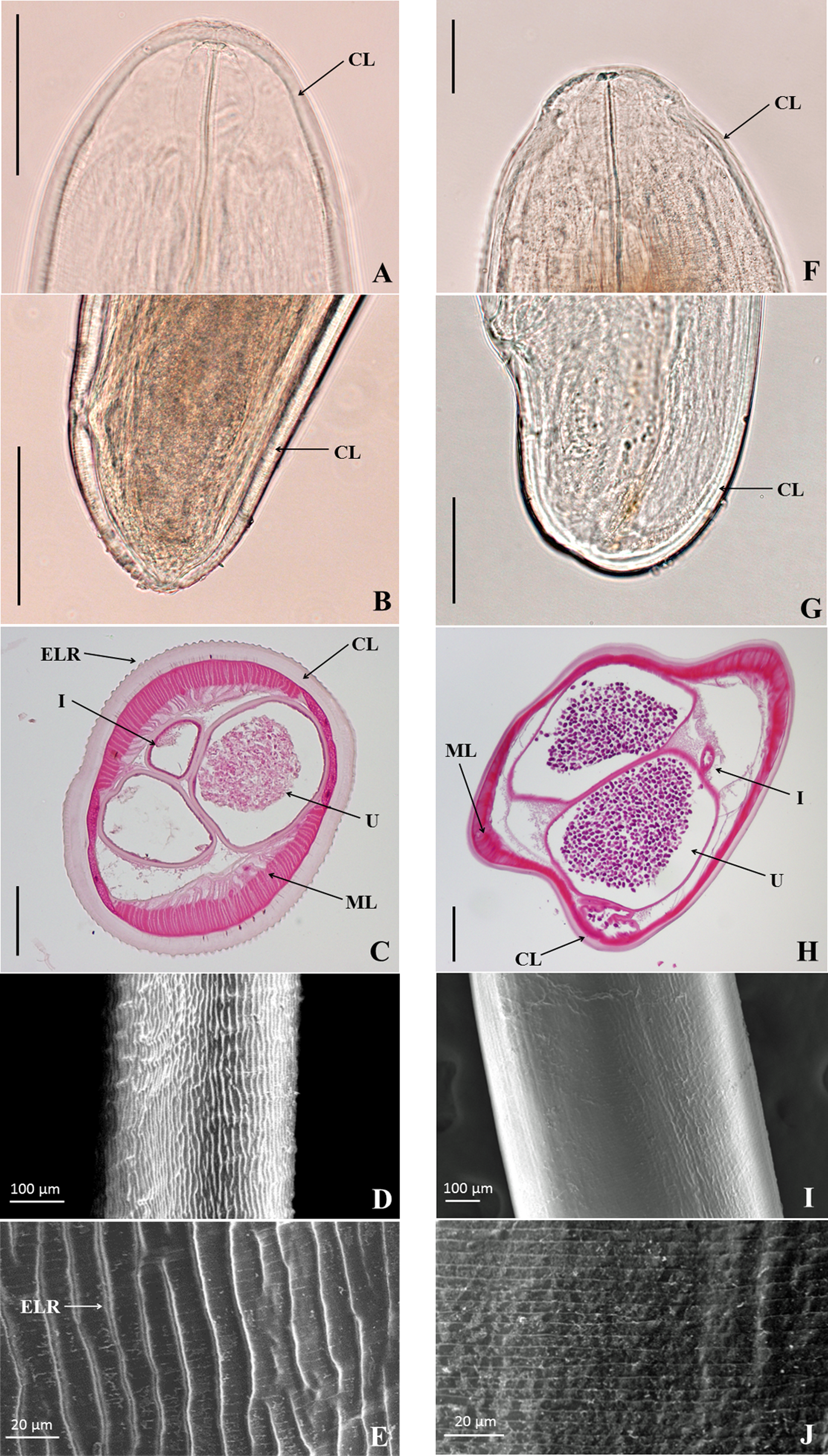
English: Dirofilaria repens removed from a subcutaneous nodule - Photos
Figure 1 of article. Morphological images of female adults of
|
| Date | |
| Source | Suzuki J, Kobayashi S, Okata U, Matsuzaki H, Mori M, Chen KR & Iwata S: Molecular analysis of Dirofilaria repens removed from a subcutaneous nodule in a Japanese woman after a tour to Europe. Parasite, 2015, 22, 2.doi:10.1051/parasite/2015002 |
| Author | Jun Suzuki, Seiki Kobayashi, Utako Okata, Hitomi Matsuzaki, Mariko Mori, Ko-Ron Chen and Satoshi Iwata |
Licensing
This file is licensed under the Creative Commons Attribution 4.0 International license.
|
 This file was published in the scientific journal Parasite. Their website states that all content of the journal including and after 2013 is published under the Creative Commons Attribution 4.0 license.
|
File history
Click on a date/time to view the file as it appeared at that time.
| Date/Time | Thumbnail | Dimensions | User | Comment | |
|---|---|---|---|---|---|
| current | 14:58, 28 January 2015 | | 1,645 × 2,894 (5.31 MB) | commons>Jeanloujustine | User created page with UploadWizard |
File usage
There are no pages that use this file.