License
Attribution 4.0 International (CC BY 4.0)
Summary
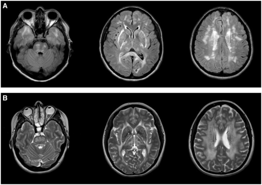
Author:David S. Lynch, Anderson Rodrigues Brandão de Paiva, Wei Jia Zhang, Enrico Bugiardini, Fernando Freua, Leandro Tavares Lucato, Lucia Inês Macedo-Souza, Rahul Lakshmanan, Justin A. Kinsella, Aine Merwick, Alexander M. Rossor, Nin Bajaj, Brian Herron, Paul McMonagle, Patrick J. Morrison, Deborah Hughes, Alan Pittman, Matilde Laurà, Mary M Reilly, Jason D Warren, Catherine J Mummery, Jonathan M. Schott, Matthew Adams, Nick C. Fox, Elaine Murphy, Indran Davagnanam, Fernando Kok, Jeremy Chataway, and Henry Houlden,Department of Molecular Neuroscience, UCL Institute of Neurology,Leonard Wolfson Experimental Neurology Centre, UCL Institute of Neurology,Neurogenetics Unit, Neurology Department, Hospital das Clínicas da Universidade de São Paulo, MRC Centre for Neuromuscular Diseases, UCL Institute of Neurology,Instituto de Radiologia, Hospital das Clínicas da Universidade de São Paulo, Centro de Estudos do Genoma Humano, Universidade de São Paulo, Lysholm Department of Neuroradiology, National Hospital for Neurology and Neurosurgery, Neurology Department, St. Vincent’s University Hospital and University College Charles Dent Metabolic Unit, National Hospital for Neurology and Neurosurgery, Chelsea and Westminster NHS Foundation Trust, Sobell Department of Motor Neuroscience and Movement Disorders, UCL Institute of Neurology, Department of Neurology, Queens Medical Centre,Department of Neuropathology, Royal Victoria Hospital, Department of Neurology, Royal Victoria Hospital, Centre for Cancer Research and Cell Biology, Queens University of Belfast, Dementia Research Centre, UCL Institute of Neurology, Department of Neuroinflammation, UCL Institute of Neurology, Neurogenetics Laboratory, National Hospital for Neurology and Neurosurgery(Openi/National Library of Medicine) Source:https://openi.nlm.nih.gov/detailedresult?img=PMC5405235_awx045f2&query=&req=4 Description:awx045-F2: CADASIL and CARASAL imaging appearance. (A) Typical imaging appearance of CADASIL in axial FLAIR MRI images. There is symmetric subcortical high signal in the anterior temporal lobes, internal and external capsules and scattered asymmetric involvement of the periventricular cerebral white matter and pons. (B) CARASAL in T2 axial images. There was no involvement of the anterior temporal lobes (left) but there was extensive involvement of the internal and external capsules, the basal ganglia and thalami (middle) and the periventricular and deep white matter (right).
.png)
.png)