File:PMC5145840 11832 2016 790 Fig18 HTML.png

From WikiProjectMed
Jump to navigation Jump to search

PMC5145840_11832_2016_790_Fig18_HTML.png(512 × 244 pixels, file size: 106 KB, MIME type: image/png)

This file is from a shared repository and may be used by other projects. The description on its file description page there is shown below.

Summary

Description
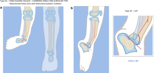
English⧼Colon⧽ Fig18: a, b Paley type 3c fibular hemimelia. The foot is in fixed equino-valgus. There is a fibrous and cartilaginous fibular anlage. There is a malunited subtalar coalition. The calcaneus is laterally translated and valgus (a). The tibial plafond is maloriented into valgus procurvatum (b). The tibial diaphysis has an antero-lateral bow. There is a leg length discrepancy. Reproduced with permission by the Paley Foundation
Date
Source https://openi.nlm.nih.gov/detailedresult?img=PMC5145840_11832_2016_790_Fig18_HTML&query=Fibular%20hemimelia&it=xg&req=4&npos=6
Author Dror Paley

Licensing

{{subst:Custom license marker added by UW}} https://creativecommons.org/licenses/by/4.0/ Attribution 4.0 International (CC BY 4.0)

This file was uploaded with UploadWizard.

File history

Click on a date/time to view the file as it appeared at that time.

Date/TimeThumbnailDimensionsUserComment
current22:46, 22 February 2023Thumbnail for version as of 22:46, 22 February 2023512 × 244 (106 KB)Ozzie10aaaaUploaded a work by Dror Paley from https://openi.nlm.nih.gov/detailedresult?img=PMC5145840_11832_2016_790_Fig18_HTML&query=Fibular%20hemimelia&it=xg&req=4&npos=6 with UploadWizard

There are no pages that use this file.