License
Attribution 4.0 International (CC BY 4.0)
Summary
Author:Paul Capewell,Christelle Cren-Travaillé, Francesco Marchesi, Pamela Johnston, Caroline Clucas, Robert A Benson, Taylor-Anne Gorman, Estefania Calvo-Alvarez, Aline Crouzols, Grégory Jouvion, Vincent Jamonneau, William Weir, M Lynn Stevenson, Kerry O'Neill, Anneli Cooper, Nono-raymond Kuispond Swar, Bruno Bucheton, Dieudonné Mumba Ngoyi, Paul Garside, Brice Rotureau, Annette MacLeod,Wellcome Trust Centre for Molecular Parasitology, University of Glasgow, College of Medical, Veterinary and Life Sciences, University of Glasgow, Henry Wellcome Building for Comparative Medical Sciences, University of Glasgow, Trypanosome Transmission Group, Trypanosome Cell Biology Unit, Department of Parasites and Insect Vectors, Institut Pasteur, Veterinary Diagnostic Services, Veterinary School, University of Glasgow, Institute of Infection, Immunology and Inflammation, University of Glasgow, Glasgow Biomedical Research Centre, University of Glasgow, Human Histopathology and Animal Models Unit, Institut Pasteur, Paris, France
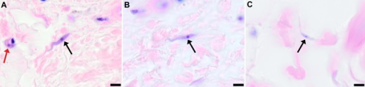
10Institut de Recherche pour le Développement, International de Baillarguet, University of Kinshasa, Kinshasa, Department of Parasitology, National Institute of Biomedical Research, Kinshasa(Openi/National Library of Medicine) Source:https://openi.nlm.nih.gov/detailedresult?img=PMC5065312_elife-17716-fig5&query=Human%20trypanosomiasis&it=xg&req=4&npos=1 Description:fig5: Extravascular localisation of trypanosomes in previously unidentified human cases of trypanosomiasisHistological sections of skin collected from previously unidentified cases of human trypanosomiasis from the Democratic Republic of Congo, showing the presence of extravascular parasites in biopsies from three individuals (A, B and C). Skin biopsies were collected as part of a national onchocerciasis screening programme that took place in the same geographic region as an active trypanosomiasis focus. Slides were stained with Giemsa and examined under oil immersion at 100x magnification. In addition to visible slender forms (black arrows) in the extravascular tissue of the skin, a clearly identifiable stumpy transmission form with typical morphology and an unattached undulating membrane is also present in the skin of one individual (red arrow in A). The scale bar represents 5 µm.DOI:http://dx.doi.org/10.7554/eLife.17716.021