No higher resolution available.
This file is from a shared repository and may be used by other projects.
The description on its file description page there is shown below.
License
Attribution-NonCommercial-ShareAlike 3.0 Unported (CC BY-NC-SA 3.0)
Summary
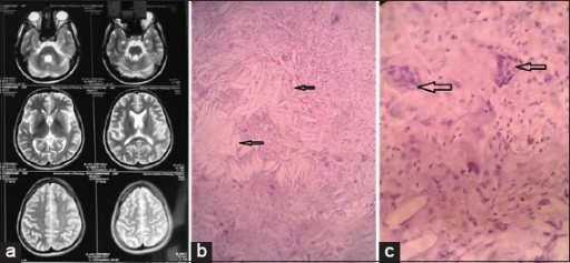
Author:Mahalakshmi Muniaswamy, Madhu Rengasamy, Ramesh Aravamuthan, and Manoharan Krishnasamy,Department of Dermatology, Madras Medical College (Openi/National Library of Medicine) Source:https://openi.nlm.nih.gov/detailedresult?img=PMC4976429_IDOJ-7-336-g002&query=cerebral%20cholesterosis&it=xg&req=4&npos=1 Description:F2: (a) MRI image of the brain shows hyperintensities in middle cerebellar region suggestive of cholesterol deposits. (b) Histopathology of thelesion with H and E stain x10 magnification shows collection of foamy histiocytes, numerous cholesterol clefts (arrows) surrounded by foreign body type of multinucleated giant cells in a background of stromal fibroblastic proliferation, consistent with tendon xanthoma. (c) Histopathology of thelesion with H and E stain x40 magnification shows collection of foamy histiocytes, numerous cholesterol clefts surrounded by foreign body type of multinucleated giant cells (arrows) in a background of stromal fibroblastic proliferation consistent with tendon xanthomas
File history
Click on a date/time to view the file as it appeared at that time.
| Date/Time | Thumbnail | Dimensions | User | Comment |
|---|
| current | 18:49, 25 October 2021 |  | 512 × 237 (269 KB) | Ozzie10aaaa | Author:Mahalakshmi Muniaswamy, Madhu Rengasamy, Ramesh Aravamuthan, and Manoharan Krishnasamy,Department of Dermatology, Madras Medical College (Openi/National Library of Medicine) Source:https://openi.nlm.nih.gov/detailedresult?img=PMC4976429_IDOJ-7-336-g002&query=cerebral%20cholesterosis&it=xg&req=4&npos=1 Description:F2: (a) MRI image of the brain shows hyperintensities in middle cerebellar region suggestive of cholesterol deposits. (b) Histopathology of thelesion with H and E stain x10 ma... |
File usage
The following page uses this file: