File:PMC4899637 cde-0008-0026-g02.png
Jump to navigation
Jump to search

Size of this preview: 263 × 598 pixels. Other resolutions: 105 × 240 pixels | 450 × 1,024 pixels.
Original file (450 × 1,024 pixels, file size: 1.11 MB, MIME type: image/png)
File history
Click on a date/time to view the file as it appeared at that time.
| Date/Time | Thumbnail | Dimensions | User | Comment | |
|---|---|---|---|---|---|
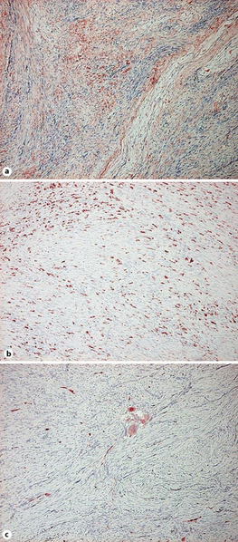
| current | 20:22, 8 March 2022 | | 450 × 1,024 (1.11 MB) | Ozzie10aaaa | Author:Shido K, Fujimura T, Kakizaki A, Furudate S, Asano M, Aiba S,Department of Dermatology, Tohoku University Graduate School of Medicine (Openi/National Library of Medicine) Source:https://openi.nlm.nih.gov/detailedresult?img=PMC4899637_cde-0008-0026-g02&query=Plexiform%20fibrohistiocytic%20tumor&it=xg&req=4&npos=9 Description:F2: Paraffin-embedded tissue samples were deparaffinized and stained with anti-POSTN antibody (a), anti-CD163 antibody (b) and anti-CD206 antibody (c). The sections... |
File usage
There are no pages that use this file.