File:PMC4899564 gr1.png
Jump to navigation
Jump to search
No higher resolution available.
PMC4899564_gr1.png (512 × 122 pixels, file size: 52 KB, MIME type: image/png)
File history
Click on a date/time to view the file as it appeared at that time.
| Date/Time | Thumbnail | Dimensions | User | Comment | |
|---|---|---|---|---|---|
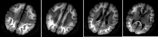
| current | 22:49, 23 December 2021 | 512 × 122 (52 KB) | Ozzie10aaaa | Author:Chang W, Gupta N, Duane D, Barnes P, Yeom K,Gundersen Lutheran Medical Foundation,Stanford University (Openi/National Library of Medicine) Source:https://openi.nlm.nih.gov/detailedresult?img=PMC4899564_gr1&query=Methylmalonic%20acidemia&it=xg&req=4&npos=2 Description:fig1: 6-day-old infant with methylmalonic acidemia. Sequential mean diffusion images derived from diffusion tensor imaging showed symmetric diffusion restriction in the subcortical white matter of bilateral cerebral hemisp... |
File usage
The following page uses this file: