File:PMC4802527 gr2.png
Jump to navigation
Jump to search
No higher resolution available.
PMC4802527_gr2.png (512 × 383 pixels, file size: 482 KB, MIME type: image/png)
File history
Click on a date/time to view the file as it appeared at that time.
| Date/Time | Thumbnail | Dimensions | User | Comment | |
|---|---|---|---|---|---|
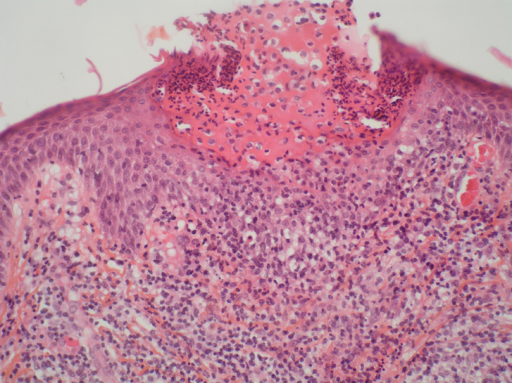
| current | 19:33, 15 April 2022 | | 512 × 383 (482 KB) | Ozzie10aaaa | Author:Ropars N, Darrieux L, Tisseau L, Safa G ,Department of Dermatology, Centre Hospitalier de Saint-Brieuc(Openi/National Library of Medicine) Source:https://openi.nlm.nih.gov/detailedresult?img=PMC4802527_gr2&query=&req=4 Description:fig2: Acute generalized exanthematous pustulosis. A skin biopsy shows a subcorneal pustule filled with neutrophils. Note the presence of necrotic keratinocytes in the epidermis. Papillary dermal edema and superficial mixed infiltrate composed of lymphocytes... |
File usage
The following page uses this file: